The MEL-14 monoclonal antibody specifically binds to CD62L (L-selectin), a 95 kDa (on neutrophils) or 74 kDa (on lymphocytes) receptor with lectin-like and Epidermal Growth Factor-like domains. In the mouse, L-selectin is detected on most thymocytes, with the highest levels of expression on an immunocompetent subset and a population of dividing progenitor cells, and on peripheral leukocytes, including subsets of B and T lymphocytes, neutrophils, monocytes, and eosinophils. This member of the selectin adhesion molecule family appears to be required for lymphocyte homing to peripheral lymph nodes and to contribute to neutrophil emigration at inflammatory sites. L-selectin is rapidly shed from lymphocytes and neutrophils upon cellular activation; metalloproteinases may mediate the release of CD62L ectodomains from the cell surface. The level of CD62L expression, along with other markers, distinguishes naive, effector, and memory T cells. L-selectin binds to sialytaed oligosaccharide determinants on high endothelial venules (HEV) in peripheral lymph nodes. In vitro studies have demonstrated that CD34, GlyCAM-1, and MAdCAM-1, all recognized by mAb MECA-79 (anti-mouse PNAd Carbohydrate Epitope, Cat. No. 553863), may be ligands for CD62L. MEL-14 mAb blocks in vitro binding of lymphocytes to peripheral lymph node HEV and inhibits in vivo lymphocyte extravasation into peripheral lymph nodes and late stages of leukocyte rolling.
商品描述
MEL-14
The MEL-14 monoclonal antibody specifically binds to CD62L (L-selectin), a 95 kDa (on neutrophils) or 74 kDa (on lymphocytes) receptor with lectin-like and Epidermal Growth Factor-like domains. In the mouse, L-selectin is detected on most thymocytes, with the highest levels of expression on an immunocompetent subset and a population of dividing progenitor cells, and on peripheral leukocytes, including subsets of B and T lymphocytes, neutrophils, monocytes, and eosinophils. This member of the selectin adhesion molecule family appears to be required for lymphocyte homing to peripheral lymph nodes and to contribute to neutrophil emigration at inflammatory sites. L-selectin is rapidly shed from lymphocytes and neutrophils upon cellular activation; metalloproteinases may mediate the release of CD62L ectodomains from the cell surface. The level of CD62L expression, along with other markers, distinguishes naive, effector, and memory T cells. L-selectin binds to sialytaed oligosaccharide determinants on high endothelial venules (HEV) in peripheral lymph nodes. In vitro studies have demonstrated that CD34, GlyCAM-1, and MAdCAM-1, all recognized by mAb MECA-79 (anti-mouse PNAd Carbohydrate Epitope, Cat. No. 553863), may be ligands for CD62L. MEL-14 mAb blocks in vitro binding of lymphocytes to peripheral lymph node HEV and inhibits in vivo lymphocyte extravasation into peripheral lymph nodes and late stages of leukocyte rolling.
同种型
Rat F344, also known as Fischer, CDF IgG2a, κ
克隆号
克隆 MEL-14 (RUO)
浓度
0.2 mg/ml
产品详情
RB705
The BD Horizon RealBlue™ 705 (RB705) Dye is part of the BD® family of blue dyes. It is a tandem fluorochrome with an excitation maximum (Ex Max) at 498-nm and an emission maximum (Em Max) at 707-nm as measured using an antibody-dye conjugate. Driven by BD® innovation, RB705 can be used on both spectral and conventional cytometers and is designed to be excited by the Blue laser (488-nm) with minimal excitation by the 561-nm Yellow-Green laser. For conventional instruments equipped with a Blue laser (488-nm), RB705 can be used as an alternative to PerCP-Cy5.5 or BB700 and we recommend using an optical filter centered near 710-nm (e.g., a 695/40 or 710/50-nm bandpass filter). For spectral instruments equipped with a Blue laser (488-nm), it can be used in conjunction with PerCP-Cy5.5. RB705 is on average brighter than PerCP-Cy5.5 and BB700, and has minimal spillover into Yellow-Green detectors.
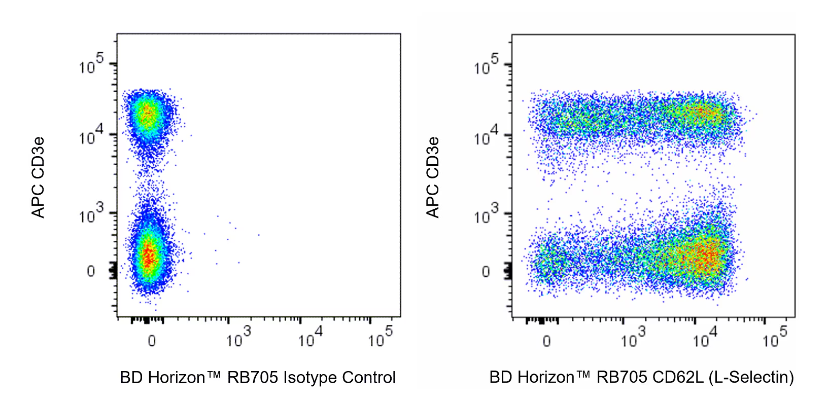
Multicolor flow cytometric analysis of CD62L (L-Selectin) expression on viable Mouse splenic leukocytes. C57BL/6 Mouse splenocytes were preincubated with Purified Rat Anti-Mouse CD16/CD32 antibody (Mouse BD Fc Block™) [Cat. No. 553141/553142]. The cells were then stained with APC Hamster Anti-Mouse CD3e (Cat. No. 553066) and with either BD Horizon™ RB705 Rat IgG2a, κ Isotype Control (Cat. No. 570262; Left Plot) or BD Horizon™ RB705 Rat Anti-Mouse CD62L (L-Selectin) antibody (Cat. No. 570282/570283; Right Plot) at 0.125 µg/test. DAPI Solution (Cat. No. 564907) was added to cells right before analysis. The bivariate pseudocolor density plot showing the correlated expression of CD62L (L-Selectin) [or Ig Isotype control staining] versus CD3e was derived from gated events with the forward and side light-scatter characteristics of viable (DAPI-negative) splenic leukocytes. Flow cytometry and data analysis were performed using a BD FACSymphony™ A5 SE Cell Analyzer System and FlowJo™ software. Data shown on this Technical Data Sheet are not lot specific.
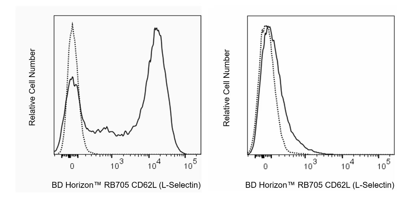
Flow cytometric analysis of CD62L (L-Selectin) expression on viable Mouse bone marrow cells. C57BL/6 mouse bone marrow cells were left untreated (Left Plot) or were cultured (1 hour) with Phorbol 12-Myristate 13-Acetate (PMA; Right Plot). The cells were preincubated with Purified Rat Anti-Mouse CD16/CD32 antibody (Mouse BD Fc Block™) [Cat. No. 553141/553142]. The cells were then stained with either BD Horizon™ RB705 Rat IgG2a, κ Isotype Control (Cat. No. 570262) or BD Horizon™ RB705 Rat Anti-Mouse CD62L (L-Selectin) antibody (Cat. No. 570282/570283) at 0.125 µg/test. DAPI (4',6-Diamidino-2-Phenylindole, Dihydrochloride) Solution (Cat. No. 564907) was added to cells right before analysis. The fluorescence histograms showing CD62L (L-Selectin) expression (or Ig Isotype control staining) were derived from gated events with the forward and side light-scatter characteristics of viable (DAPI-negative) cells. Flow cytometry and data analysis were performed using a BD FACSymphony™ A5 SE Cell Analyzer System and FlowJo™ software. Data shown on this Technical Data Sheet are not lot specific.


 全部商品分类


 下载产品说明书
 用小程序,查商品更便捷


 收藏
收藏
 对比
对比 咨询
咨询
