


 下载产品说明书 下载SDS
 用小程序,查商品更便捷


 收藏
收藏
 对比
对比 咨询
咨询
Ala19-Asp412
Accession # P50454
Scientific Data
 View Larger
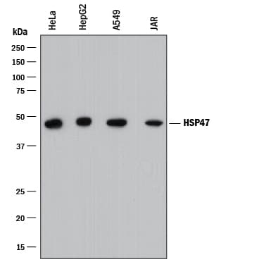
Detection of Human HSP47 by Western Blot. Western blot shows lysates of HeLa human cervical epithelial carcinoma cell line, HepG2 human hepatocellular carcinoma cell line, A549 human lung carcinoma cell line, and JAR human choriocarcinoma cell line. PVDF membrane was probed with 0.1 µg/mL of Mouse Anti-Human HSP47 Monoclonal Antibody (Catalog # MAB91662) followed by HRP-conjugated Anti-Mouse IgG Secondary Antibody (Catalog # HAF018). A specific band was detected for HSP47 at approximately 47 kDa (as indicated). This experiment was conducted under reducing conditions and using Immunoblot Buffer Group 1.
.") View Larger
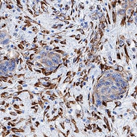
View LargerHSP47 in Human Breast Cancer Tissue. HSP47 was detected in immersion fixed paraffin-embedded sections of human breast cancer tissue using Mouse Anti-Human HSP47 Monoclonal Antibody (Catalog # MAB91662) at 25 µg/mL overnight at 4 °C. Tissue was stained using the Anti-Mouse HRP-DAB Cell & Tissue Staining Kit (brown; Catalog # CTS002) and counterstained with hematoxylin (blue). Specific staining was localized to cytoplasm. View our protocol for Chromogenic IHC Staining of Paraffin-embedded Tissue Sections.
 View Larger
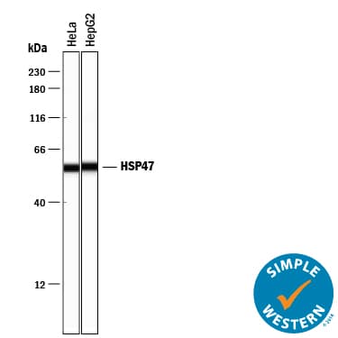
Detection of Human HSP47 by Simple WesternTM. Simple Western lane view shows lysates of HeLa human cervical epithelial carcinoma cell line and HepG2 human hepatocellular carcinoma cell line, loaded at 0.2 mg/mL. A specific band was detected for HSP47 at approximately 57 kDa (as indicated) using 1 µg/mL of Mouse Anti-Human HSP47 Monoclonal Antibody (Catalog # MAB91662). This experiment was conducted under reducing conditions and using the 12-230 kDa separation system.
Human HSP47 Antibody Summary
Ala19-Asp412
Accession # P50454
Applications
Please Note: Optimal dilutions should be determined by each laboratory for each application. General Protocols are available in the Technical Information section on our website.
Background: HSP47
Heat Shock Protein 47 (HSP47), also known as Serpin-H1/CBP1/CBP2, is localized to endoplasmic reticulum (ER), where it is a collagen-specific molecular chaperone. In the ER, HSP47 interacts with and stabilizes correctly-folded procollagen. Nucleotide polymorphisms may be associated with preterm birth and Osteogenesis Imperfecta type X. Serpin-H1 is up-regulated in several cancers including squamous cell carcinoma, breast and prostate carcinomas. Expression in tumors drives malignant growth and invasion by enhancing deposition of extracellular matrix proteins.
- Christiansen HE, et al, (2010) Am. J. Hum. Genet. 86:3892.
- Tasab M, et al, (2000) EMBO J. 19:22043.
- Kwon YJ, et al, (2009) Oncol Res. 18:1414.
- Zhu J, et al, (2015) Cancer Res. 75:15805.
- Nese N, et al, (2010) Anal Quant Cytol Histol. 32:90
Preparation and Storage
- 12 months from date of receipt, -20 to -70 °C as supplied.
- 1 month, 2 to 8 °C under sterile conditions after reconstitution.
- 6 months, -20 to -70 °C under sterile conditions after reconstitution.
参考图片
HSP47 in Human Breast Cancer Tissue. HSP47 was detected in immersion fixed paraffin-embedded sections of human breast cancer tissue using Mouse Anti-Human HSP47 Monoclonal Antibody (Catalog # MAB91662) at 25 µg/mL overnight at 4 °C. Tissue was stained using the Anti-Mouse HRP-DAB Cell & Tissue Staining Kit (brown; Catalog # CTS002) and counterstained with hematoxylin (blue). Specific staining was localized to cytoplasm. View our protocol for Chromogenic IHC Staining of Paraffin-embedded Tissue Sections.
Detection of Human HSP47 by Western Blot. Western blot shows lysates of HeLa human cervical epithelial carcinoma cell line, HepG2 human hepatocellular carcinoma cell line, A549 human lung carcinoma cell line, and JAR human choriocarcinoma cell line. PVDF membrane was probed with 0.1 µg/mL of Mouse Anti-Human HSP47 Monoclonal Antibody (Catalog # MAB91662) followed by HRP-conjugated Anti-Mouse IgG Secondary Antibody (Catalog # HAF018). A specific band was detected for HSP47 at approximately 47 kDa (as indicated). This experiment was conducted under reducing conditions and using Immunoblot Buffer Group 1.
Detection of Human HSP47 by Simple WesternTM. Simple Western lane view shows lysates of HeLa human cervical epithelial carcinoma cell line and HepG2 human hepatocellular carcinoma cell line, loaded at 0.2 mg/mL. A specific band was detected for HSP47 at approximately 57 kDa (as indicated) using 1 µg/mL of Mouse Anti-Human HSP47 Monoclonal Antibody (Catalog # MAB91662). This experiment was conducted under reducing conditions and using the 12-230 kDa separation system.

 危险品化学品经营许可证(不带存储) 许可证编号:沪(杨)应急管危经许[2022]202944(QY)
  营业执照(三证合一)