


 全部商品分类


SIRPalpha/SHPS1 (D6I3M) Rabbit mAb

 下载产品说明书 下载COA 下载SDS
 用小程序,查商品更便捷


 收藏
收藏
 对比
对比 咨询
咨询Monoclonal antibody is produced by immunizing animals with a synthetic peptide corresponding to residues surrounding Pro413 of human SIRPα/SHPS1 protein.

Product Usage Information
| Application | Dilution |
|---|---|
| Western Blotting | 1:1000 |
| IHC Leica Bond | 1:50 - 1:200 |
| Immunohistochemistry (Paraffin) | 1:50 - 1:100 |
| Immunofluorescence (Immunocytochemistry) | 1:50 - 1:100 |
| Flow Cytometry (Fixed/Permeabilized) | 1:50 - 1:200 |
Specificity/Sensitivity
Species Reactivity:
Human, Mouse, Rat, Monkey
Supplied in 10 mM sodium HEPES (pH 7.5), 150 mM NaCl, 100 µg/ml BSA, 50% glycerol and less than 0.02% sodium azide. Store at –20°C. Do not aliquot the antibody.
For a carrier free (BSA and azide free) version of this product see product #47027.
参考图片
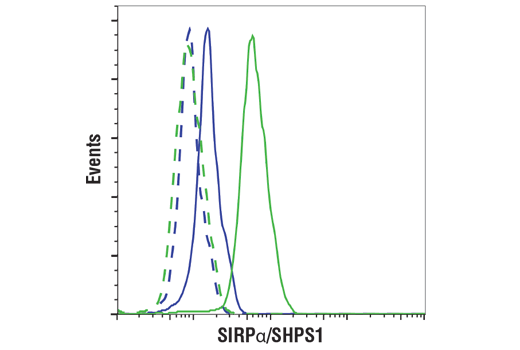
Flow cytometric analysis of fixed and permeabilized Jurkat cells (blue, negative) and U937 cells (green, positive) using SIRPα/SHPS1 (D6I3M) Rabbit mAb (solid lines) or a concentration-matched Rabbit (DA1E) mAb IgG XP® Isotype Control #3900 (dashed lines). Anti-rabbit IgG (H+L), F(ab')2 Fragment (Alexa Fluor® 488 Conjugate) #4412 was used as a secondary antibody.
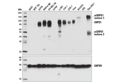
Western blot analysis of extracts from various cell lines using SIRPα/SHPS1 (D6I3M) Rabbit mAb (upper) and GAPDH (D16H11) XP® Rabbit mAb #5174 (lower).
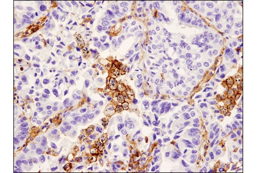
Immunohistochemical analysis of paraffin-embedded breast ductal carcinoma using SIRPa/SHPS1 (D6I3M) Rabbit mAb performed on the Leica® BOND™ Rx.
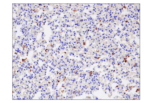
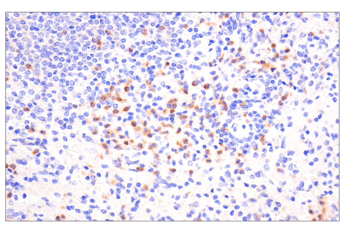
Immunohistochemical analysis of paraffin-embedded mouse lung using SIRPa/SHPS1 (D6I3M) Rabbit mAb performed on the Leica® BOND™ Rx.
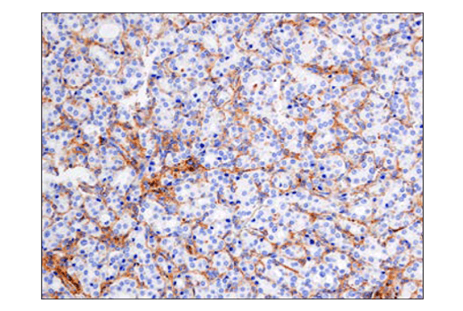
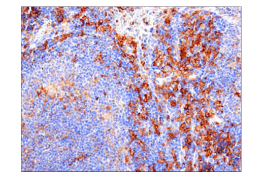
Immunohistochemical analysis of paraffin-embedded mouse spleen using SIRPa/SHPS1 (D6I3M) Rabbit mAb performed on the Leica® BOND™ Rx.
Immunohistochemical analysis of paraffin-embedded human lung carcinoma using SIRPα/SHPS1 (D6I3M) Rabbit mAb.
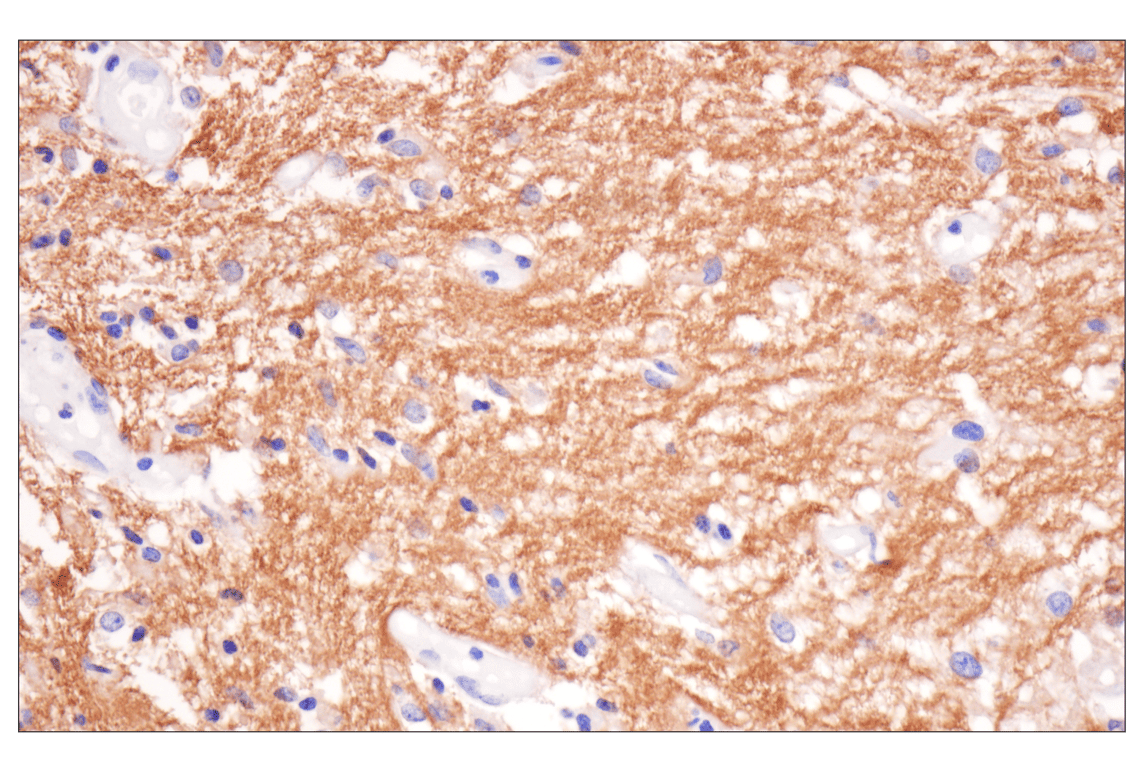
Immunohistochemical analysis of paraffin-embedded normal human brain using SIRPa/SHPS1 (D6I3M) Rabbit mAb.
Immunohistochemical analysis of paraffin-embedded normal human spleen using SIRPa/SHPS1 (D6I3M) Rabbit mAb.
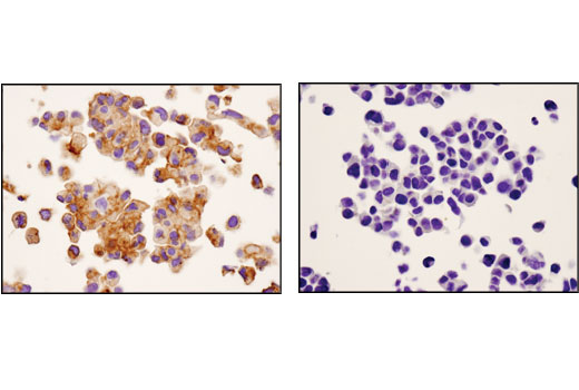
Immunohistochemical analysis of paraffin-embedded ACHN (left) or MCF7 (right) cell pellets using SIRPα/SHPS1 (D6I3M) Rabbit mAb.
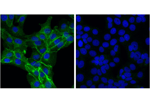
Confocal immunofluorescent analysis of ACHN (positive, left) and MCF7 (low expression, right) cells, using SIRPα/SHPS1 (D6I3M) Rabbit mAb (green). Blue pseudocolor= DRAQ5® #4084 (fluorescent DNA dye).