


 全部商品分类


SMAD2/3 Antibody Sampler Kit

 下载产品说明书 下载SDS
 用小程序,查商品更便捷


 收藏
收藏
 对比
对比 咨询
咨询
The SMAD2/3 Antibody Sampler Kit provides an economical means of detecting target proteins of the TGF-β signaling pathway. The kit includes enough antibody to perform two western blots with each primary antibody.
参考图片
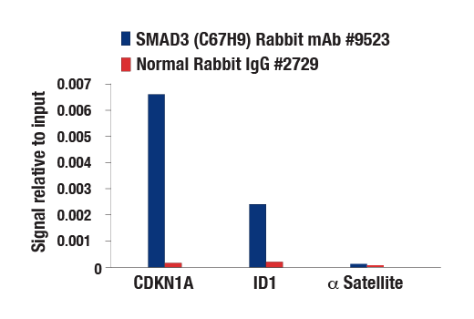
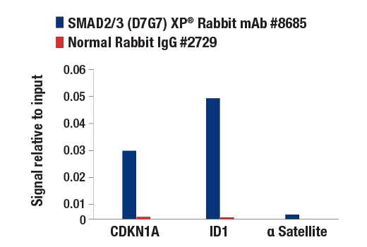
Chromatin immunoprecipitations were performed with cross-linked chromatin from HaCaT cells treated with Human TGF-β3 #3706 (7 ng/ml) for 1 h and either SMAD3 (C67H9) Rabbit mAb or Normal Rabbit IgG #2729 using SimpleChIP® Plus Enzymatic Chromatin IP Kit (Magnetic Beads) #9005. The enriched DNA was quantified by real-time PCR using SimpleChIP® Human CDKN1A Intron 1 Primers #4669, SimpleChIP® Human ID1 Promoter Primers #5139, and SimpleChIP® Human α Satellite Repeat Primers #4486. The amount of immunoprecipitated DNA in each sample is represented as signal relative to the total amount of input chromatin, which is equivalent to one.
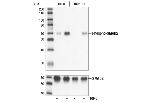
Western blot analysis of extracts from untreated or TGF-beta treated HeLa and NIH/3T3 cells, using Phospho-SMAD2 (Ser465/467) (138D4) Rabbit mAb (upper), or SMAD2 Antibody #3102 (lower).
Simple Western™ analysis of lysates (1.0 mg/mL) from serum starved HT-1080 cells treated with hTGF-β3 (10 ng/mL, 30 min) using Phospho-SMAD2 (Ser465/467) (138D4) Rabbit mAb #3108. The virtual lane view (left) shows a single target band (as indicated) at 1:50 and 1:250 dilutions of primary antibody. The corresponding electropherogram view (right) plots chemiluminescence by molecular weight along the capillary at 1:50 (blue line) and 1:250 (green line) dilutions of primary antibody. This experiment was performed under reducing conditions on the Jess™ Simple Western instrument from ProteinSimple, a BioTechne brand, using the 12-230 kDa separation module.
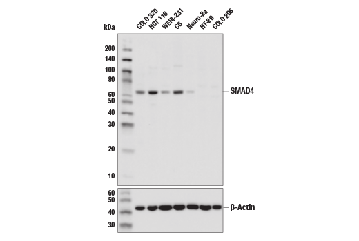
Western blot analysis of extracts from various cell lines using SMAD4 (D3M6U) Rabbit mAb (upper) and β-Actin (D6A8) Rabbit mAb #8457 (lower). HT-29 and COLO 205 are SMAD4-null mutant cell lines, confirming specificity of the antibody.
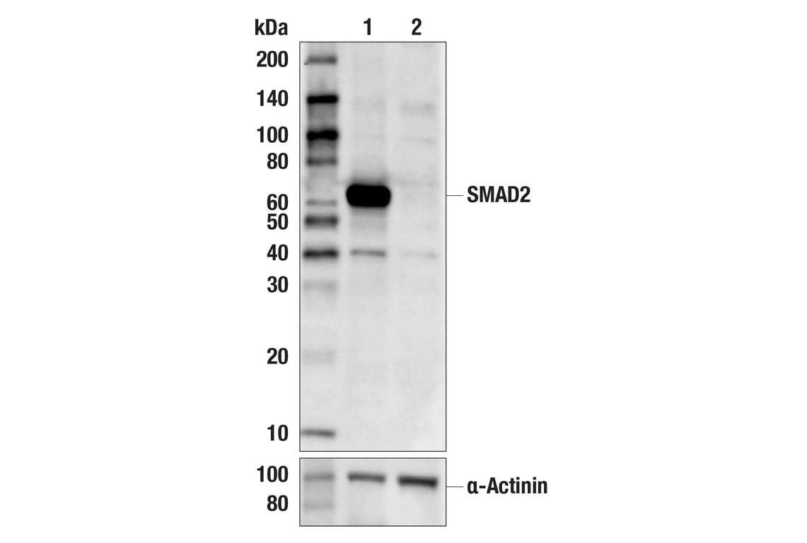
Western blot analysis of extracts from HeLa cells (lane 1) or SMAD2 knock-out cells (lane 2) using Smad2 (D43B4) XP® Rabbit mAb #5339 (upper), and α-Actinin (D6F6) XP® Rabbit mAb #6487 (lower). The absence of signal in the SMAD2 knock-out HeLa cells confirms specificity of the antibody for SMAD2.
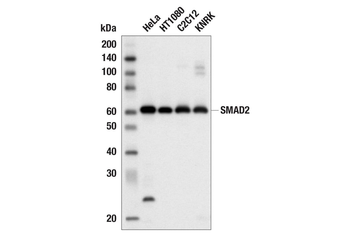
Western blot analysis of extracts from various cell lines using Smad2 (D43B4) XP® Rabbit mAb.
Confocal immunofluorescent analysis of NIH/3T3 cells, serum-starved (left) or treated with hTGF-β3 #8425 (right), using Smad2 (D43B4) XP® Rabbit mAb (green). Actin filaments have been labeled with DY-554 phalloidin (red).
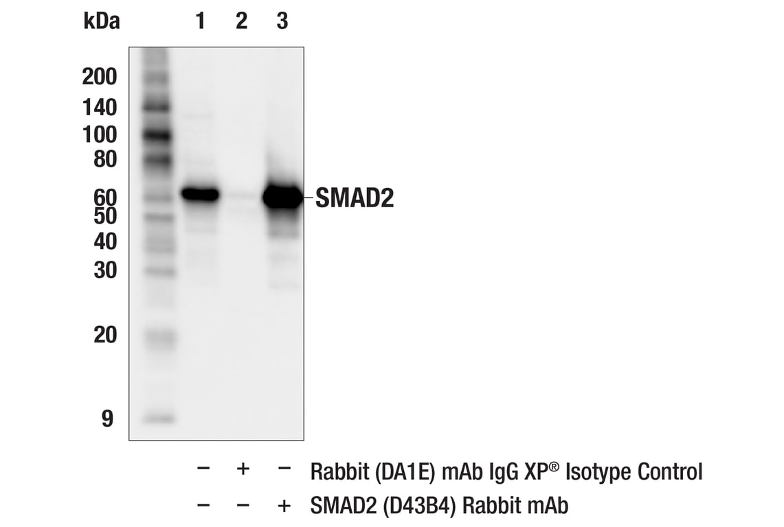
Immunoprecipitation of Smad2 from HeLa cell extracts. Lane 1 is 10% input, lane 2 is Rabbit (DA1E) mAb IgG XP® Isotype Control #3900, and lane 3 is SMAD2 (D43B4) XP® Rabbit mAb. Western blot analysis was performed using SMAD2 (L16D3) Mouse mAb #3103. Anti-mouse IgG, HRP-linked Antibody #7076 was used as a secondary antibody.
After the primary antibody is bound to the target protein, a complex with HRP-linked secondary antibody is formed. The LumiGLO® is added and emits light during enzyme catalyzed decomposition.
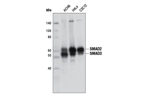
Western blot analysis of extracts from HeLa and ACHN cells using SMAD2/3 (D7G7) XP® Rabbit mAb.
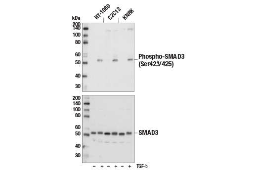
Western blot analysis of extracts from HT-1080, C2C12, or KNRK cells, untreated (-) or treated with TGF-β (10 ng/ml, 30 min; +), using Phospho-SMAD3 (Ser423/425) (C25A9) Rabbit mAb #9520 (upper) or total SMAD3 (C67H9) Rabbit mAb #9523 (lower).
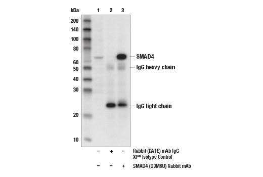
Immunoprecipitation of SMAD4 protein from HCT 116 cell extracts. Lane 1 is 10% input, lane 2 is Rabbit (DA1E) mAb IgG XP® Isotype Control #3900, and lane 3 is SMAD4 (D3M6U) Rabbit mAb. Western blot analysis was performed using SMAD4 (D3M6U) Rabbit mAb.
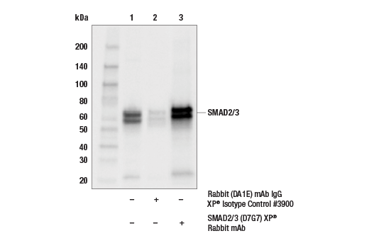
Immunoprecipitation of SMAD2/3 protein from HCT116 extracts. Lane 1 is 10% input, lane 2 is Rabbit (DA1E) mAb IgG XP® Isotype Control #3900, and lane 3 is SMAD2/3 (D7G7) XP®. Western blot analysis was performed using SMAD2/3 (D7G7) XP®. Mouse Anti-rabbit IgG (Conformation Specific) (L27A9) mAb #3678 was used as a secondary antibody.
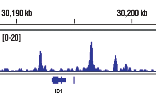
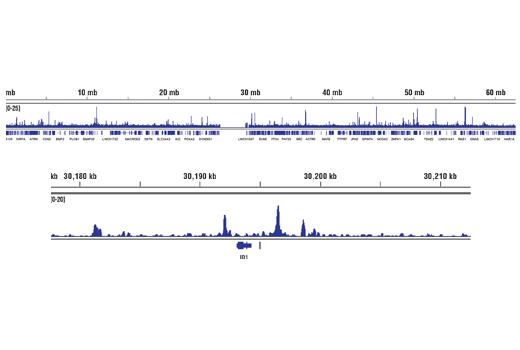
Chromatin immunoprecipitations were performed with cross-linked chromatin from HaCaT cells treated with TGF-β1 #8915 (7 ng/mL, 1 hr) and SMAD4 (D3M6U) Rabbit mAb, using SimpleChIP® Plus Enzymatic Chromatin IP Kit (Magnetic Beads) #9005. DNA Libraries were prepared using SimpleChIP® ChIP-seq DNA Library Prep Kit for Illumina® #56795. The figure shows binding across ID1, a known target gene of SMAD4 (see additional figure containing ChIP-qPCR data).
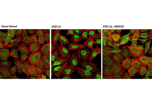
Confocal immunofluorescent analysis of HeLa cells, serum starved (left), treated with hTGF-β3 #8425 (100 ng/ml, 30 min, center), or treated with hTGF-β3 and SB43152 (10 ug/mL, 1 hr, right), using SMAD2/3 (D7G7) XP® Rabbit mAb (green). Actin filaments were labeled with DY-554 phalloidin (red).
Chromatin immunoprecipitations were performed with cross-linked chromatin from HaCaT cells treated with Human TGF-β3 #3706 (7 ng/ml) for 1 h and either Phospho-SMAD3 (Ser423/425) (C25A9) Rabbit mAb #9520 or Normal Rabbit IgG #2729 using SimpleChIP® Enzymatic Chromatin IP Kit (Magnetic Beads) #9003. The enriched DNA was quantified by real-time PCR using SimpleChIP® Human CDKN1A Intron 1 Primers #4669, SimpleChIP® Human ID1 Promoter Primers #5139, human c-Myc intron 1 primers, and SimpleChIP® Human α Satellite Repeat Primers #4486. The amount of immunoprecipitated DNA in each sample is represented as signal relative to the total amount of input chromatin, which is equivalent to one.
Chromatin immunoprecipitations were performed with cross-linked chromatin from HaCaT cells treated with TGF-β1 #8915 (7 ng/mL, 1 hr) and SMAD4 (D3M6U) Rabbit mAb, using SimpleChIP® Plus Enzymatic Chromatin IP Kit (Magnetic Beads) #9005. DNA Libraries were prepared using SimpleChIP® ChIP-seq DNA Library Prep Kit for Illumina® #56795. The figure shows binding across chromosome 20 (upper), including ID1 (lower), a known target gene of SMAD4 (see additional figure containing ChIP-qPCR data).
Flow cytometric analysis of HeLa cells using SMAD2 (D43B4) XP® Rabbit mAb (solid line) compared to concentration-matched Rabbit (DA1E) mAb IgG XP® Isotype Control #3900 (dashed line). Anti-rabbit IgG (H+L), F(ab')2 Fragment (Alexa Fluor® 488 Conjugate) #4412 was used as a secondary antibody.
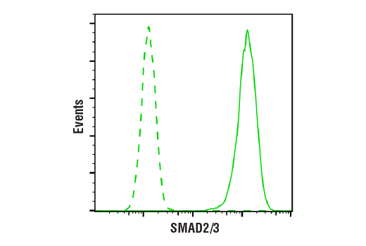
Flow cytometric analysis of HeLa cells using SMAD2/3 (D7G7) XP® Rabbit mAb (solid line) compared to concentration-matched Rabbit (DA1E) mAb IgG XP® Isotype Control #3900 (dashed line). Anti-rabbit IgG (H+L), F(ab')2 Fragment (Alexa Fluor® 488 Conjugate) #4412 was used as a secondary control.
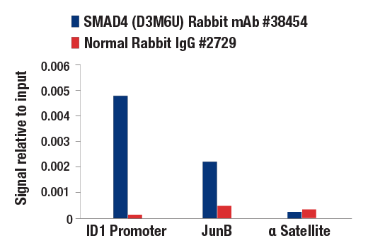
Chromatin immunoprecipitations were performed with cross-linked chromatin from HaCaT cells treated with TGF-β1 #8915 (7 ng/mL, 1 hr) and either SMAD4 (D3M6U) Rabbit mAb or Normal Rabbit IgG #2729 using SimpleChIP® Enzymatic Chromatin IP Kit (Magnetic Beads) #9003. The enriched DNA was quantified by real-time PCR using SimpleChIP® Human ID1 Promoter Primers #5139, human JunB promoter primers, and SimpleChIP® Human α Satellite Repeat Primers #4486. The amount of immunoprecipitated DNA in each sample is represented as signal relative to the total amount of input chromatin (equivalent to one).
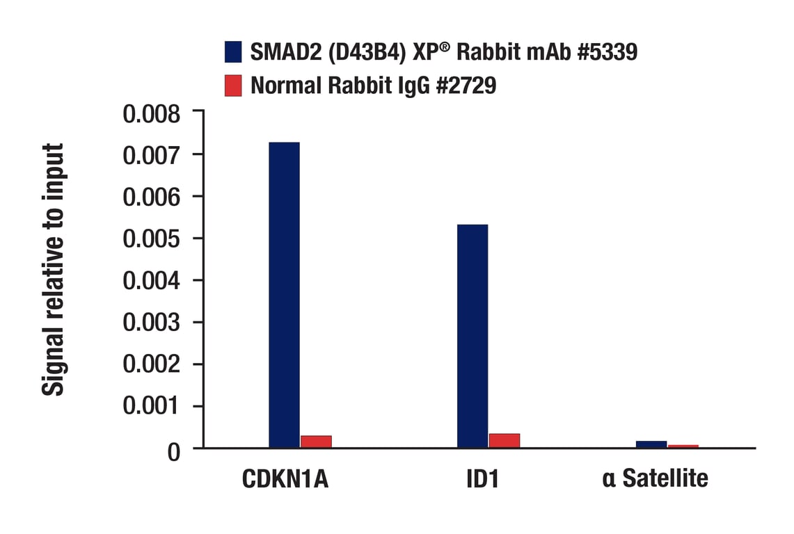
Chromatin immunoprecipitations were performed with cross-linked chromatin from HaCaT cells treated with Human TGF-β3 #8425 (7 ng/ml) for 1 h and either Smad2 (D43B4) XP® Rabbit mAb #5339 or Normal Rabbit IgG #2729 using SimpleChIP® Plus Enzymatic Chromatin IP Kit (Magnetic Beads) #9005. The enriched DNA was quantified by real-time PCR using SimpleChIP® Human CDKN1A Intron 1 Primers #4669, SimpleChIP® Human ID1 Promoter Primers #5139, and SimpleChIP® Human α Satellite Repeat Primers #4486. The amount of immunoprecipitated DNA in each sample is represented as signal relative to the total amount of input chromatin, which is equivalent to one.
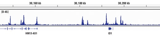
Chromatin immunoprecipitations were performed with cross-linked chromatin from HaCaT cells treated with hTGF-β3 #8425 (7 ng/ml, 1 hr) and SMAD2/3 (D7G7) XP® Rabbit mAb, using SimpleChIP® Enzymatic Chromatin IP Kit (Magnetic Beads) #9003. DNA Libraries were prepared using DNA Library Prep Kit for Illumina® (ChIP-seq, CUT&RUN) #56795. The figure shows binding across ID1, a known target gene of SMAD2/3 (see additional figure containing ChIP-qPCR data). For additional ChIP-seq tracks, please download the product datasheet.
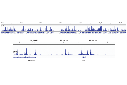
Chromatin immunoprecipitations were performed with cross-linked chromatin from HaCaT cells treated with hTGF-β3 #8425 (7 ng/ml, 1 hr) and SMAD2/3 (D7G7) XP® Rabbit mAb, using SimpleChIP® Enzymatic Chromatin IP Kit (Magnetic Beads) #9003. DNA Libraries were prepared using DNA Library Prep Kit for Illumina® (ChIP-seq, CUT&RUN) #56795. The figure shows binding across chromosome 20 (upper), including ID1 (lower), a known target gene of SMAD2/3 (see additional figure containing ChIP-qPCR data).
Chromatin immunoprecipitations were performed with cross-linked chromatin from HaCaT cells treated with hTGF-β3 #8425 (7 ng/ml, 1 hr) and either SMAD2/3 (D7G7) XP® Rabbit mAb or Normal Rabbit IgG #2729 using SimpleChIP® Enzymatic Chromatin IP Kit (Magnetic Beads) #9003. The enriched DNA was quantified by real-time PCR using SimpleChIP® Human CDKN1A Intron 1 Primers #4669, SimpleChIP® Human ID1 Promoter Primers #5139, and SimpleChIP® Human α Satellite Repeat Primers #4486. The amount of immunoprecipitated DNA in each sample is represented as signal relative to the total amount of input chromatin, which is equivalent to one.
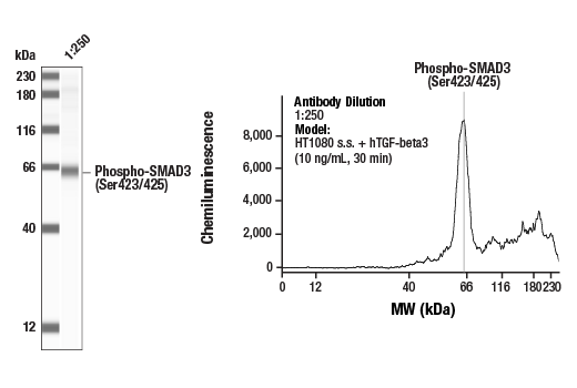
Simple Western™ analysis of lysates (1 mg/mL) from serum-starved HT1080 cells treated with hTGF-beta3 (10 ng/mL, 30 min) using Phospho-SMAD3 (Ser423/425) (C25A9) Rabbit mAb #9520. The virtual lane view (left) shows the target band (as indicated) at a 1:250 dilution of primary antibody. The corresponding electropherogram view (right) plots chemiluminescence by molecular weight along the capillary at a 1:250 dilution (gray line) of primary antibody. This experiment was performed under reducing conditions on the Jess™ Simple Western instrument from ProteinSimple, a BioTechne brand, using the 12-230 kDa separation module.
Western blot analysis of extracts from control HeLa cells (lane 1) or HeLa cells with an apparent in-frame truncation mutation in the gene encoding SMAD3 (lane 2) using SMAD3 (C67H9) Rabbit mAb #9523 (upper) or β-actin (D6A8) Rabbit mAb #8457 (lower). The change in SMAD3 molecular weight in the mutated HeLa cells is consistent with an in-frame deletion.
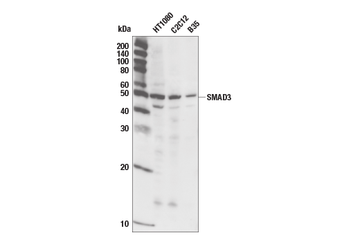
Western blot analysis of extracts from HT1080 (human), C2C12 (mouse) and B35 (rat) using SMAD3 (C67H9) Rabbit mAb.
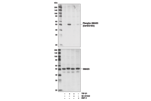
Western blot analysis of extracts from HT1080 cells, treated with TGF-β1, TGFR inhibitor SB-431542 or BMP-2, using Phospho-SMAD3 (Ser423/425) (C25A9) Rabbit mAb #9520 (upper) or total SMAD3 (C67H9) Rabbit mAb #9523 (lower).
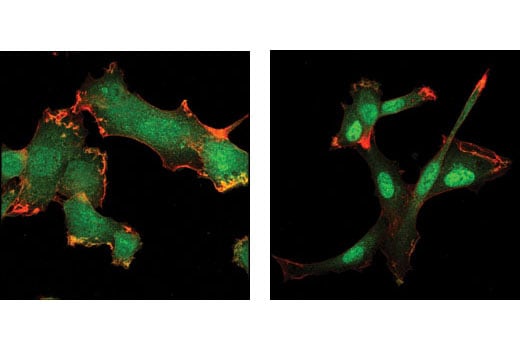
Confocal immunofluorescent analysis of HT1080 cells, untreated (left) or TGFβ-treated (right), using SMAD3 (C67H9) Rabbit mAb (green). Actin filaments have been labeled with Alexa Fluor® 555 phalloidin (red).
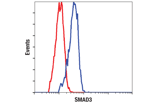
Flow cytometric analysis of HT-1080 cells using SMAD3 (C67H9) Rabbit mAb #9523 (blue) compared to a nonspecific negative control antibody (red).
Chromatin immunoprecipitations were performed with cross-linked chromatin from HaCaT cells treated with Human TGF-β3 #3706 (7 ng/ml) for 1 h and SMAD3 (C67H9) Rabbit mAb, using SimpleChIP® Plus Enzymatic Chromatin IP Kit (Magnetic Beads) #9005. DNA Libraries were prepared using DNA Library Prep Kit for Illumina® (ChIP-seq, CUT&RUN) #56795. The figure shows binding across CDKN1A, a known target gene of SMAD3 (see additional figure containing ChIP-qPCR data).
Chromatin immunoprecipitations were performed with cross-linked chromatin from HaCaT cells treated with Human TGF-β3 #3706 (7 ng/ml) for 1 h and SMAD3 (C67H9) Rabbit mAb, using SimpleChIP® Plus Enzymatic Chromatin IP Kit (Magnetic Beads) #9005. DNA Libraries were prepared using DNA Library Prep Kit for Illumina® (ChIP-seq, CUT&RUN) #56795. The figure shows binding across chromosome 6 (upper), including CDKN1A (lower), a known target gene of SMAD3 (see additional figure containing ChIP-qPCR data).