


 全部商品分类
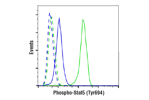


Phospho-Stat5 (Tyr694) (D47E7) XP ® Rabbit mAb

 下载产品说明书 下载COA 下载SDS
 用小程序,查商品更便捷


 收藏
收藏
 对比
对比 咨询
咨询
Monoclonal antibody is produced by immunizing animals with a synthetic peptide corresponding to residues surrounding Tyr694 of human Stat5a protein.
Product Usage Information
| Application | Dilution |
|---|---|
| Western Blotting | 1:1000 |
| Simple Western™ | 1:10 - 1:50 |
| Immunofluorescence (Immunocytochemistry) | 1:100 |
| Flow Cytometry (Fixed/Permeabilized) | 1:100 - 1:400 |
Specificity/Sensitivity
Species Reactivity:
Human, Mouse
Supplied in 10 mM sodium HEPES (pH 7.5), 150 mM NaCl, 100 µg/ml BSA, 50% glycerol and less than 0.02% sodium azide. Store at –20°C. Do not aliquot the antibody.
For a carrier free (BSA and azide free) version of this product see product #72712.
参考图片
Flow cytometric analysis of TF-1 cells, untreated (blue, low) or treated with hGM-CSF #8922 (50 ng/ml, 15 min; green, positive) using Phospho-Stat5 (Tyr694) (D47E7) XP® Rabbit mAb (solid lines) or concentration-matched Rabbit (DA1E) mAb IgG XP® Isotype Control #3900 (dashed lines). Anti-rabbit IgG (H+L), F(ab')2 Fragment (Alexa Fluor® 488 Conjugate) #4412 was used as a secondary antibody.
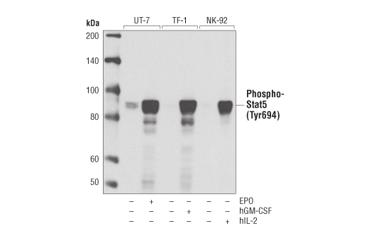
Western blot analysis of extracts from UT-7 cells, untreated or treated with erythropoietin (EPO; 3 units/ml for 5 min), TF-1 cells, untreated or treated with Human Granulocyte Macrophage Colony Stimulating Factor #8922 (hGM-CSF; 100 ng/ml for 10 min), and NK-92 cells, untreated or treated with Human Interleukin-2 #8907 (hIL-2; 100 ng/ml for 10 min), using Phospho-Stat5 (Tyr694) (D47E7) XP® Rabbit mAb.
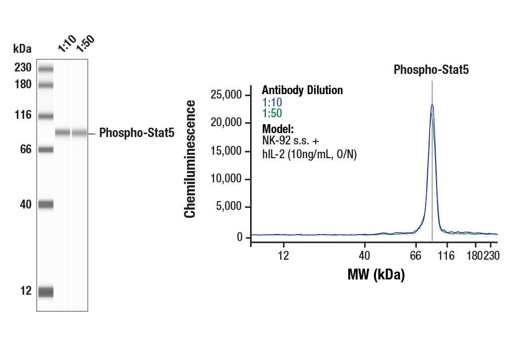
Simple Western™ analysis of lysates (1 mg/mL) from serum-starved NK-92 cells treated with hIL-2 (10 ng/mL, O/N) using Phospho-Stat5 (Tyr694) (D47E7) XP® Rabbit mAb #4322. The virtual lane view (left) shows the target band (as indicated) at 1:10 and 1:50 dilutions of primary antibody. The corresponding electropherogram view (right) plots chemiluminescence by molecular weight along the capillary at 1:10 (blue line) and 1:50 (green line) dilutions of primary antibody. This experiment was performed under reducing conditions on the Jess™ Simple Western instrument from ProteinSimple, a BioTechne brand, using the 12-230 kDa separation module.
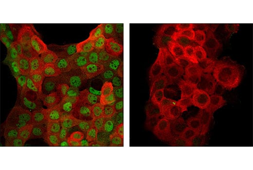
Confocal immunofluorescent analysis of A-431 cells, EGF-treated (left) or untreated (right), using Phospho-Stat5 (Tyr694) XP®(D47E7) Rabbit mAb (green) and Pan-Keratin (C11) Mouse mAb #4545 (red).