


 全部商品分类
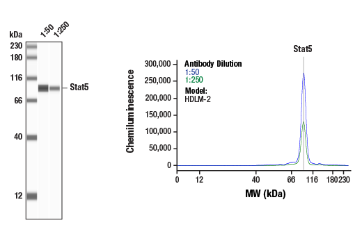


Stat Antibody Sampler Kit

 下载产品说明书 下载SDS
 用小程序,查商品更便捷


 收藏
收藏
 对比
对比 咨询
咨询
The Stat Antibody Sampler Kit provides an economical means to examine multiple Stat proteins: Stat1, Stat3, Stat5 and Stat6. The kit contains enough primary and secondary antibodies to perform two Western blot experiments.
参考图片
Simple Western™ analysis of lysates (0.1 mg/mL) from HDLM-2 cells using Stat5 (D2O6Y) Rabbit mAb #94205. The virtual lane view (left) shows the target band (as indicated) at 1:50 and 1:250 dilutions of primary antibody. The corresponding electropherogram view (right) plots chemiluminescence by molecular weight along the capillary at 1:50 (blue line) and 1:250 (green line) dilutions of primary antibody. This experiment was performed under reducing conditions on the Jess™ Simple Western instrument from ProteinSimple, a BioTechne brand, using the 12-230 kDa separation module.
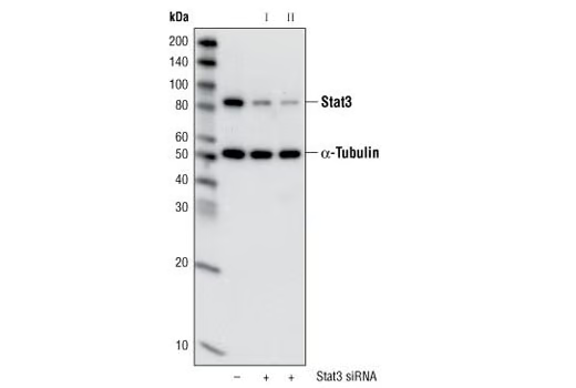
Western blot analysis of extracts from HeLa cells, transfected with 100 nM SignalSilence® Control siRNA (Unconjugated) #6568 (-), SignalSilence® Stat3 siRNA I (+) or SignalSilence® Stat3 siRNA II #6582 (+), using Stat3 (79D7) Rabbit mAb #4904 and α-Tubulin (11H10) Rabbit mAb #2125. The Stat3 (79D7) Rabbit mAb confirms silencing of Stat3 expression, while the α-Tubulin (11H10) Rabbit mAb is used to control for loading and specificity of Stat3 siRNA.
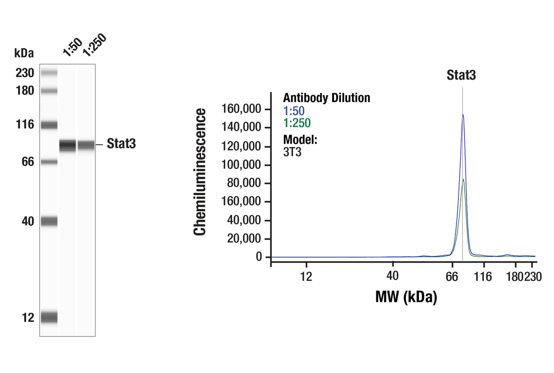
Simple WesternTM analysis of lysates (0.1 mg/mL) from 3T3 cells using Stat3 (79D7) Rabbit mAb #4904. The virtual lane view (left) shows the target band (as indicated) at 1:50 and 1:250 dilutions of primary antibody. The corresponding electropherogram view (right) plots chemiluminescence by molecular weight along the capillary at 1:50 (blue line) and 1:250 (green line) dilutions of primary antibody. This experiment was performed under reducing conditions on the JessTM Simple Western instrument from ProteinSimple, a BioTechne brand, using the 12-230 kDa separation module.
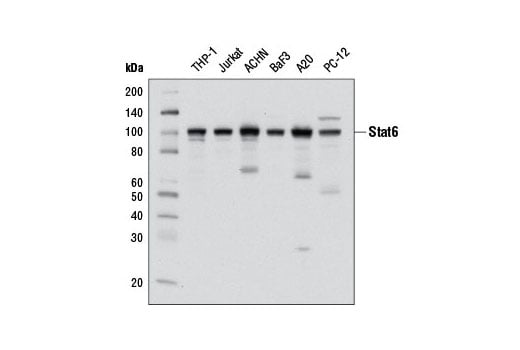
Western blot analysis of extracts from various cell lines using Stat6 (D3H4) Rabbit mAb.
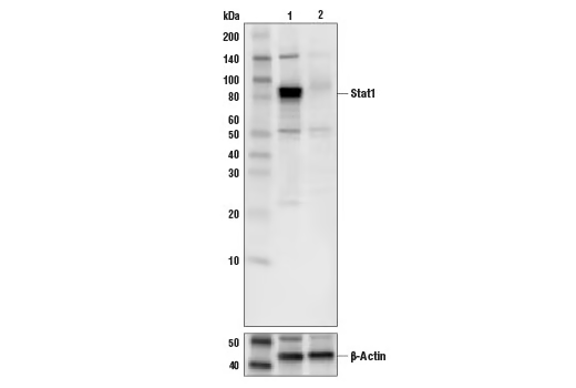
Western blot analysis of extracts from A549 cells (lane 1) or STAT1 knock-out cells (lane 2) using Stat1 Antibody #9172 (upper), and β-actin (D6A8) Rabbit mAb #8457 (lower). The absence of signal in the STAT1 knock-out A549 cells confirms specificity of the antibody for STAT1.
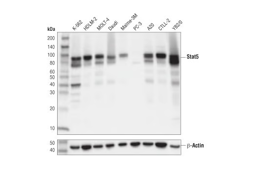
Western blot analysis of extracts from various cell lines using Stat5 (D2O6Y) Rabbit mAb (upper) and β-Actin (D6A8) Rabbit mAb #8457 (lower).
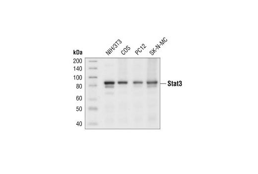
Western blot analysis of extracts from various cell lines using Stat3 (79D7) Rabbit mAb.
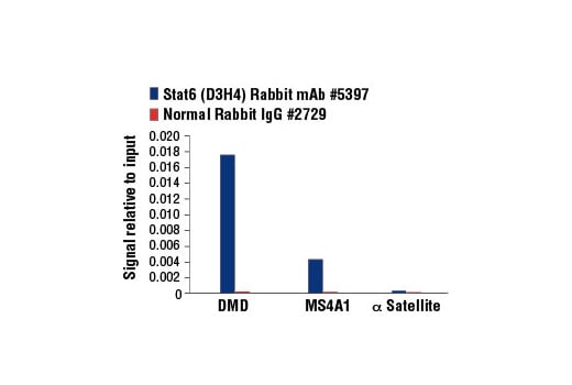
Chromatin immunoprecipitations were performed with cross-linked chromatin from Ramos cells starved overnight then treated with IL-4 (100 ng/ml, 30 min) and either Stat6 (D3H4) Rabbit mAb or Normal Rabbit IgG #2729 using SimpleChIP® Enzymatic Chromatin IP Kit (Magnetic Beads) #9003. The enriched DNA was quantified by real-time PCR using SimpleChIP® Human DMD Intron 2 Primers #7710, human MS4A1 promoter primers, and SimpleChIP® Human α Satellite Repeat Primers #4486. The amount of immunoprecipitated DNA in each sample is represented as signal relative to the total amount of input chromatin, which is equivalent to one.
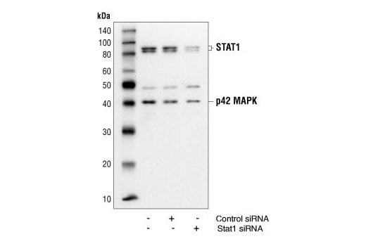
Western blot analysis of extracts from HeLa cells 48 hours following mock transfection, transfection with non-targeted (control) siRNA or transfection with Stat1 siRNA. Stat1 was detected using Stat1 Antibody #9172, and p42 was detected using p42 MAPK Antibody #9108. The Stat1 Antibody confirms silencing of Stat1 expression, and the p42 MAPK Antibody is used to control for protein loading and siRNA specificity.
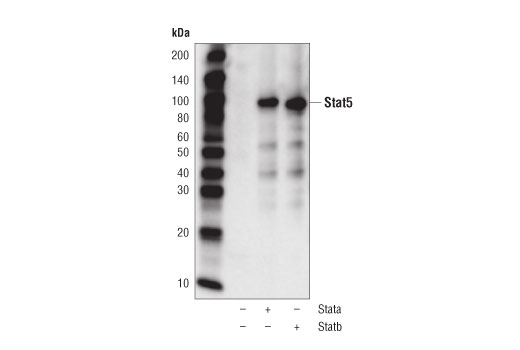
Western blot analysis of PC-3 cells, mock transfected (-) or transfected with constructs expressing full-length human Stat5a (+) or Stat5b (+) using Stat5 (D2O6Y) Rabbit mAb.
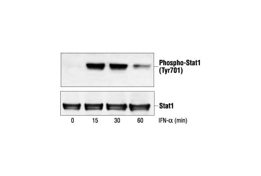
Western blot analysis of extracts from SK-MEL-28 cells, untreated or IFN-alpha-treated (100 ng/ml), using Phospho-Stat1 (Tyr701) Antibody #9171 (upper) or Stat1 Antibody (lower).
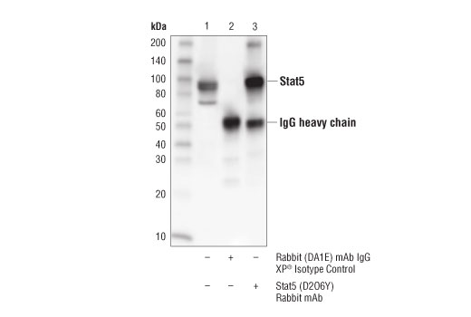
Immunoprecipitation of Stat5 from K-562 cell extracts. Lane 1 is 10% input, lane 2 is precipitated with Rabbit (DA1E) mAb IgG XP® Isotype Control #3900, and lane 3 is Stat5 (D2O6Y) Rabbit mAb. Western blot was performed using Stat5 (D2O6Y) Rabbit mAb.
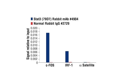
Chromatin immunoprecipitations were performed with cross-linked chromatin from Hep G2 cells treated with IL-6 (100 ng/ml) for 30 minutes, and either Stat3 (79D7) Rabbit mAb or Normal Rabbit IgG #2729 using SimpleChIP® Enzymatic Chromatin IP Kit (Magnetic Beads) #9003. The enriched DNA was quantified by real-time PCR using human IRF-1 promoter primers, SimpleChIP® Human c-Fos Promoter Primers #4663, and SimpleChIP® Human α Satellite Repeat Primers #4486. The amount of immunoprecipitated DNA in each sample is represented as signal relative to the total amount of input chromatin, which is equivalent to one.
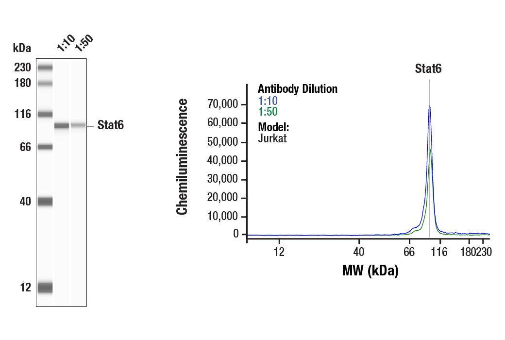
Simple Western™ analysis of lysates (0.1 mg/mL) from Jurkat cells using Stat6 (D3H4) Rabbit mAb #5397. The virtual lane view (left) shows the target band (as indicated) at 1:10 and 1:50 dilutions of primary antibody. The corresponding electropherogram view (right) plots chemiluminescence by molecular weight along the capillary at 1:10 (blue line) and 1:50 (green line) dilutions of primary antibody. This experiment was performed under reducing conditions on the Jess™ Simple Western instrument from ProteinSimple, a BioTechne brand, using the 12 – 230 kDa separation module.
After the primary antibody is bound to the target protein, a complex with HRP-linked secondary antibody is formed. The LumiGLO® is added and emits light during enzyme catalyzed decomposition.
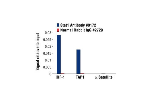
Chromatin immunoprecipitations were performed with cross-linked chromatin from HT-1080 cells treated with IFN-γ (50 ng/ml) for 30 minutes and either Stat1 Antibody or Normal Rabbit IgG #2729 using SimpleChIP® Enzymatic Chromatin IP Kit (Magnetic Beads) #9003. The enriched DNA was quantified by real-time PCR using human IRF-1 promoter primers, SimpleChIP® Human TAP1 Promoter Primers #5148, and SimpleChIP® Human α Satellite Repeat Primers #4486. The amount of immunoprecipitated DNA in each sample is represented as signal relative to the total amount of input chromatin, which is equivalent to one.
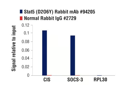
Chromatin immunoprecipitations were performed with cross-linked chromatin from BaF3 cells starved of IL-3 for 6 hours followed by induction with IL-3 for 45 minutes, and either Stat5 (D2O6Y) Rabbit mAb or Normal Rabbit IgG #2729 using SimpleChIP® Enzymatic Chromatin IP Kit (Magnetic Beads) #9003. The enriched DNA was quantified by real-time PCR using SimpleChIP® Mouse CIS Intron 1 Primers #5131, mouse SOCS-3 promoter primers, and SimpleChIP® Mouse RPL30 Intron 2 Primers #7015. The amount of immunoprecipitated DNA in each sample is represented as signal relative to the total amount of input chromatin, which is equivalent to one.