


 全部商品分类
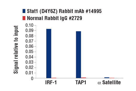


Stat1 (D4Y6Z) Rabbit mAb

 下载产品说明书 下载COA 下载SDS
 用小程序,查商品更便捷


 收藏
收藏
 对比
对比 咨询
咨询
Monoclonal antibody is produced by immunizing animals with recombinant protein specific to a region near the carboxy terminus of human Stat1 protein.
Product Usage Information
For optimal ChIP results, use 10 μl of antibody and 10 μg of chromatin (approximately 4 x 10⁶ cells) per IP. This antibody has been validated using SimpleChIP® Enzymatic Chromatin IP Kits.
| Application | Dilution |
|---|---|
| Western Blotting | 1:1000 |
| Immunoprecipitation | 1:100 |
| Immunohistochemistry (Paraffin) | 1:400 - 1:1600 |
| Immunofluorescence (Frozen) | 1:400 - 1:1600 |
| Immunofluorescence (Immunocytochemistry) | 1:400 - 1:1600 |
| Chromatin IP | 1:50 |
| Chromatin IP-seq | 1:50 |
Specificity/Sensitivity
Species Reactivity:
Human, Mouse, Rat
Supplied in 10 mM sodium HEPES (pH 7.5), 150 mM NaCl, 100 µg/ml BSA, 50% glycerol and less than 0.02% sodium azide. Store at –20°C. Do not aliquot the antibody.
For a carrier free (BSA and azide free) version of this product see product #84689.
参考图片
Chromatin immunoprecipitations were performed with cross-linked chromatin from HT-1080 cells treated with Human Interferon-γ (hIFN-γ) #8901 (50 ng/ml, 30 min) and either Stat1 (D4Y6Z) Rabbit mAb or Normal Rabbit IgG #2729 using SimpleChIP® Enzymatic Chromatin IP Kit (Magnetic Beads) #9003. The enriched DNA was quantified by real-time PCR using human IRF-1 promoter primers, SimpleChIP® Human TAP1 Promoter Primers #5148, and SimpleChIP® Human α Satellite Repeat Primers #4486. The amount of immunoprecipitated DNA in each sample is represented as signal relative to the total amount of input chromatin, which is equivalent to one.
Western blot analysis of extracts from various cell lines using Stat1 (D4Y6Z) Rabbit mAb.
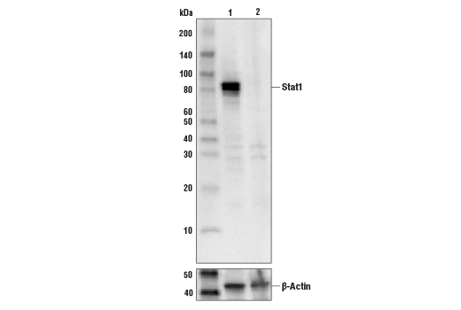
Western blot analysis of extracts from A549 cells (lane 1) or STAT1 knock-out cells (lane 2) using Stat1 (D4Y6Z) Rabbit mAb #14995 (upper), and β-actin (D6A8) Rabbit mAb #8457 (lower). The absence of signal in the STAT1 knock-out A549 cells confirms specificity of the antibody for STAT1.
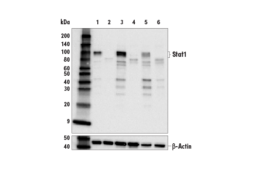
Western blot analysis of extracts from whole lung from wild type 129S6/SvEvTac mice (model #129SVE-F) (lane 1), or Stat1 knock-out mice (model #2045-F) (lane 2), whole spleen extracts from wild type 129S6/SvEvTac mice (lane 3), or Stat1 knock-out mice (lane 4), whole liver extracts from wild type 129S6/SvEvTac mice (lane 5), or Stat1 knock-out mice (lane 6) using Stat1 (D4Y6Z) Rabbit mAb #14995 (upper) and β-Actin (D6A8) Rabbit mAb #8457 (lower). The truncated protein observed in the knock-out tissues is the result of a targeted homozygous mutation in the Stat1 gene (4), and confirms the specificity of the antibody for Stat1. Mice from Taconic Biosciences, Inc.
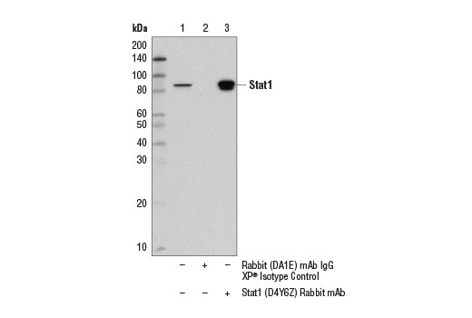
Immunoprecipitation of Stat1 from MCF7 cell extracts using Rabbit (DA1E) mAb IgG XP® Isotype Control #3900 (lane 2) or Stat1 (D4Y6Z) Rabbit mAb (lane 3). Lane 1 is 10% input. Western blot was performed using Stat1 (9H2) Mouse mAb #9176.
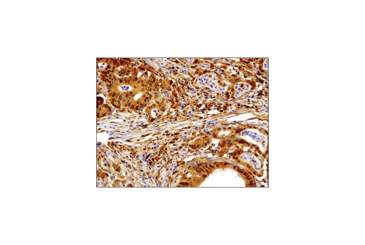
Immunohistochemical analysis of paraffin-embedded human colon carcinoma using Stat1 (D4Y6Z) Rabbit mAb.
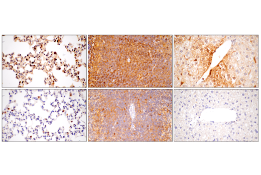
Immunohistochemical analysis of paraffin-embedded lung (left), spleen (middle), and liver (right) from wild type 129S6/SvEvTac mouse (model #129SVE-F) (top) or Stat1 knock-out mouse (model #2045-F) (bottom) using Stat1 (D4Y6Z) Rabbit mAb. The targeted mutation in the KO animal results in a reduction of expression and truncation of the protein(4). Mice from Taconic Biosciences, Inc.
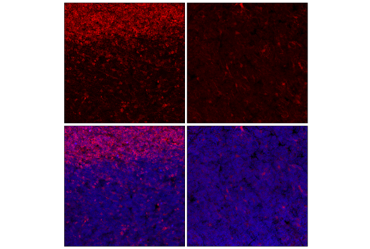
Confocal immunofluorescent analysis of fixed, frozen thymus from wild type 129S6/SvEvTac mouse (model #129SVE-F) (left) or Stat1 knock-out mouse (model #2045-F) (right) using Stat1 (D4Y6Z) Rabbit mAb #14995 (red) and DAPI #4083 (blue). The targeted mutation in the KO animal results in a reduction of expression and truncation of the protein(4). Mice from Taconic Biosciences, Inc.
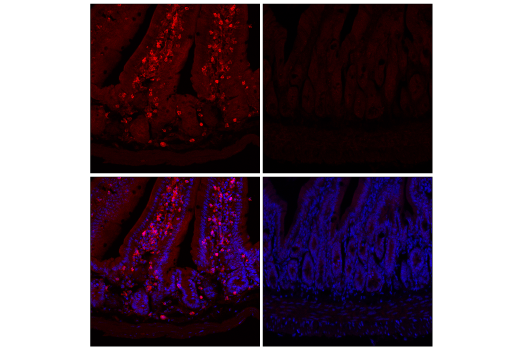
Confocal immunofluorescent analysis of fixed, frozen small intestine from wild type 129S6/SvEvTac mouse (model #129SVE-F) (left) or Stat1 knock-out mouse (model #2045-F) (right) using Stat1 (D4Y6Z) Rabbit mAb #14995 (red) and DAPI #4083 (blue). The targeted mutation in the KO animal results in a reduction of expression and truncation of the protein(4). Mice from Taconic Biosciences, Inc.
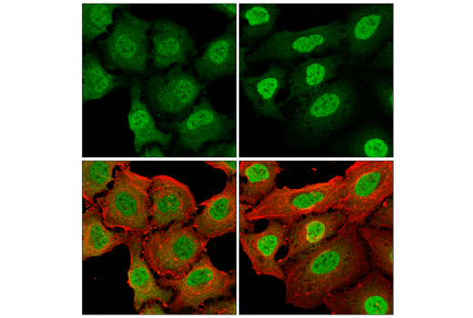
Confocal immunofluorescent analysis of HeLa cells either serum-starved overnight (left) or serum-starved overnight and treated with Human Interferon-α1 (hIFN-α1) #8927 (1,000 units/ml, 30 min; right) using Stat1 (D4Y6Z) Rabbit mAb #14995 (green) and β-Actin (8H10D10) Mouse mAb #3700 (red).