


 全部商品分类


StemLight™ Pluripotency Antibody Kit

 下载产品说明书 下载SDS
 用小程序,查商品更便捷


 收藏
收藏
 对比
对比 咨询
咨询
The StemLight® Pluripotency Antibody Kit contains a panel of antibodies for the detection of proteins that are specifically expressed in human pluripotent cells. The kit can be used to track the pluripotent potential of human embryonic stem (ES) or induced pluripotent (iPS) cells. The loss of these markers indicates a loss of pluripotency or differentiation of the culture. The kit components are pre-optimized for parallel use in immunofluorescent analysis. Enough reagents are provided for 100 assays based on a working volume of 100 µl.
参考图片
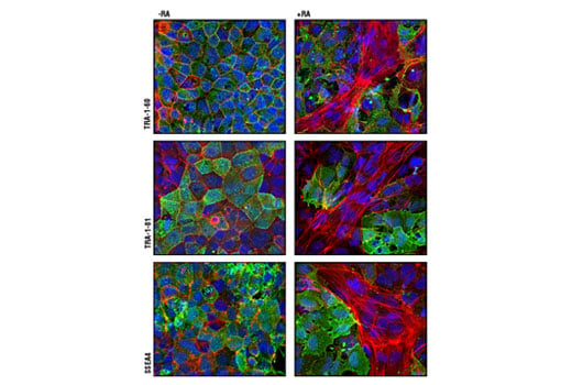
Confocal immunofluorescent analysis of NTERA-2 cells, untreated (left panel) or retinoic acid-treated (10 µM all-trans RA for 5 days) (right panel), using TRA-1-60(S) (TRA-1-60(S)) Mouse mAb (green, upper), TRA-1-81 (TRA-1-81) Mouse mAb (green, middle) and SSEA4 (MC813) Mouse mAb (green, lower). Actin filaments have been labeled with DY-554 phalloidin (red). Blue pseudocolor = DRAQ5® #4084 (fluorescent DNA dye). Note the loss of pluripotency markers (green) as cells differentiate along the neuronal lineage with retinoic acid treatment.
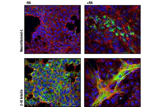
Confocal immunofluorescent analysis of NTERA-2 cells, untreated (left panel) or retinoic acid-treated (10 µM all-trans RA for 5 days) (right panel), using Neurofilament-L (C28E10) Rabbit mAb #2837 (green, upper), and β3-Tubulin (TU-20) Mouse mAb #4466 (green, lower). Actin filaments have been labeled with DY-554 phalloidin (red). Blue pseudocolor = DRAQ5® #4084 (fluorescent DNA dye). Note the appearance of neuronal markers and structures as cells differentiate along the neuronal lineage with retinoic acid treatment.
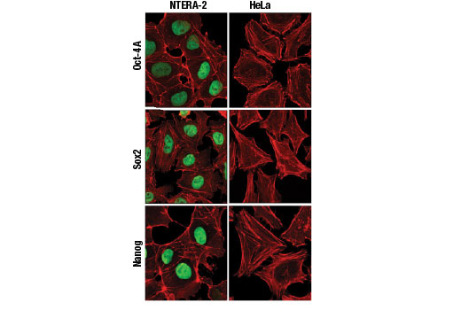
Confocal immunofluorescent analysis of NTERA-2 (left) and HeLa cells (right) using Oct-4A (C30A3) Rabbit mAb (green, upper), Sox2 (D6D9) XP® Rabbit mAb (green, middle) and Nanog (D73G4) XP® Rabbit mAb (green, lower).
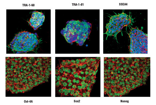
Projected confocal z-stack of human iPS cells using TRA-1-60(S) (TRA-1-60(S)) Mouse mAb (green, upper left), TRA-1-81 (TRA-1-81) Mouse mAb (green, upper middle), SSEA4 (MC813) Mouse mAb (green, upper right), Oct-4A (C30A3) Rabbit mAb (green, lower left), Sox2 (D6D9) XP® Rabbit mAb (green, lower middle) and Nanog (D73G4) XP® Rabbit mAb (green, lower right). Actin filaments were labeled with DY-554 phalloidin (red). Blue pseudocolor = DRAQ5® #4084 (fluorescent DNA dye).