Note: The panel is 50 test size. Extra material for each component for an additional 10 tests is provided for instrument set up purposes.
Multicolor immunofluorescent staining followed by flow cytometric analysis and sorting has led to the phenotypic and functional characterization of multiple peripheral T cell subsets. Three major T cell subsets have been well characterized, i.e., naïve, memory and effector T cells. Naïve T cells have a relatively homogenous cell surface phenotype. They express high levels of CD45RA and the CD197 (CCR7 chemokine receptor). Effector T cells coexpress variable to low levels of CD45RA and no CD197 whereas memory T cells coexpress variable to low levels of CD45RA and high levels of CD197. The Human Naive/Memory T Cell Panel contains fluorescent antibodies (each optimized at 5 μl per test) that are specific for the cell surface antigens: CD45RA, CD197, CD4 and CD3. The channel for detecting Phycoerythrin or Phycoerythrin protein-tandem conjugated proteins has been left open for inclusion of other markers. The panel was designed to standardize the multicolor staining and flow cytometric characterization of the three major CD4+ T cell subsets that arise as a consequence of development or clonal expansion and differentiation driven by antigenic stimulation (eg, in response to allergens, infectious disease or vaccination).
克隆号
(RUO)
BD化合物表
描述
数量/尺寸
零件号
EntrezGene ID
Alexa Fluor® 647 Mouse Anti-Human CD197 (CCR7)
60 Tests (1 ea)
51-9007097
N/A
APC-H7 Mouse Anti-Human CD3
60 Tests (1 ea)
51-9007098
N/A
PerCP-Cy™5.5 Mouse Anti-Human CD4
60 Tests (1 ea)
51-9007099
N/A
FITC Mouse Anti-Human CD45RA
60 Tests (1 ea)
51-9007100
N/A
应用
实验应用
Flow cytometry (Routinely Tested)
反应种属
Human (QC Testing)
目标/特异性
Naive, memory, effector kit
文献
文献
研发参考(5)
1. Appay V, van Lier RA, Sallusto F, Roederer M. Phenotype and function of human T lymphocyte subsets: consensus and issues. Cytometry A. 2008; 73(11):975-983. (Biology).
2. Kallies A. Distinct regulation of effector and memory T-cell differentiation. Immunol Cell Biol. 2008; 86(4):325-332. (Biology).
3. Sallusto F, Geginat J, Lanzavecchia A. Central memory and effector memory T cell subsets: function, generation, and maintenance. Annu Rev Immunol. 2004; 22:745-763. (Biology).
4. Saule P, Trauet J, Dutriez V, Lekeux V, Dessaint JP, Labalette M. Accumulation of memory T cells from childhood to old age: central and effector memory cells in CD4(+) versus effector memory and terminally differentiated memory cells in CD8(+) compartment. 2006; 127(3):274-281. (Biology).
5. Seder RA, Darrah PA, Roederer M. T-cell quality in memory and protection: implications for vaccine design. Nat Rev Immunol. 2008; 8(4):247-258. (Biology).
参考图片
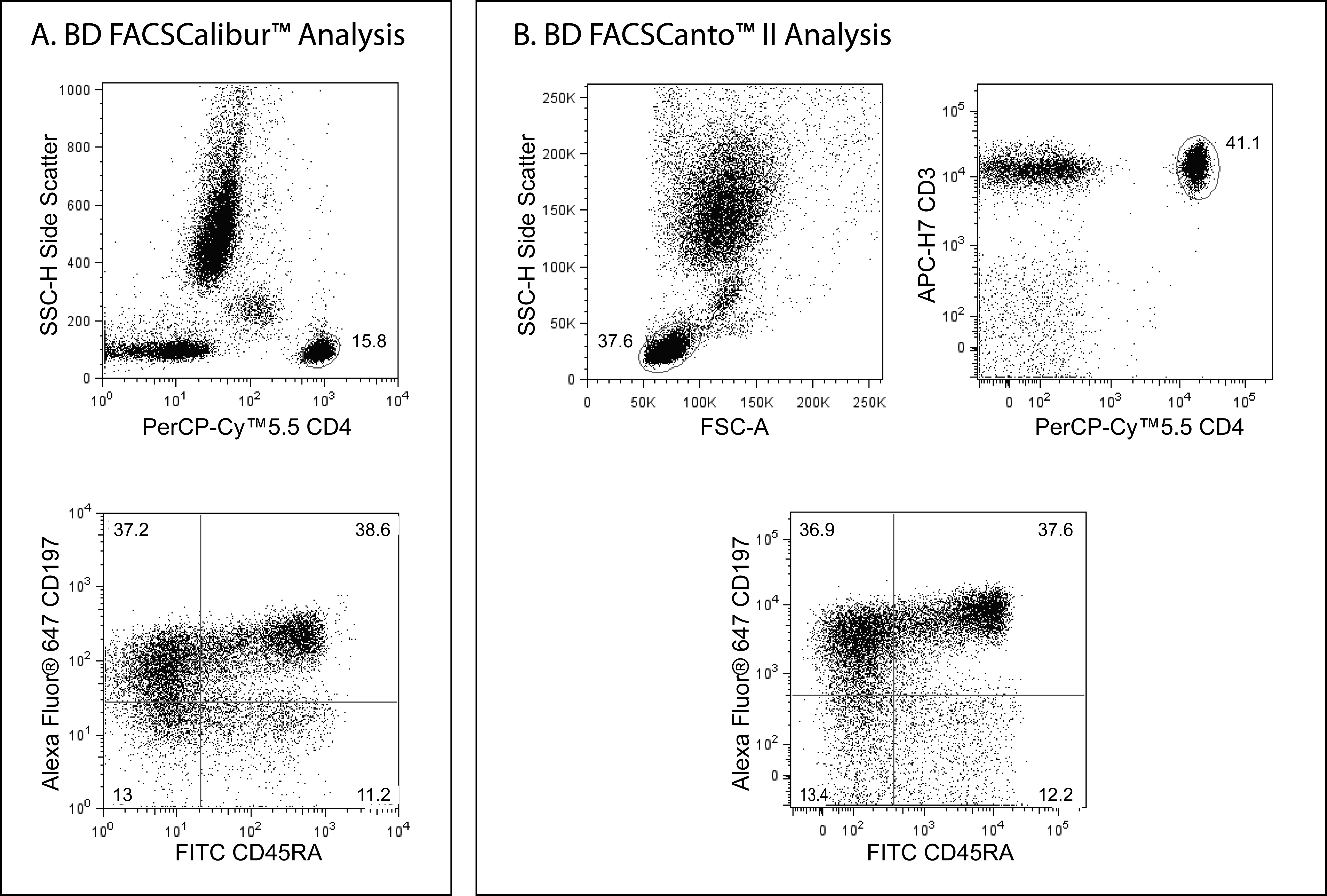
Multicolor staining and flow cytometric analysis of peripheral blood naïve and memory CD4+ T-cells using BD FACSCalibur™ and BD FACSCanto™ Cytometry Systems. Whole blood was stained with a combination of PerCP-Cy™5.5 Mouse anti-Human CD4, FITC Mouse anti-Human CD45RA, and Alexa Fluor® 647 Mouse anti-Human CD197 (CCR7) antibodies for analysis with the BD FACSCalibur™ Cytometer System, or with the addition of APC-H7 Mouse anti-Human CD3 antibody, for analysis with the BD FACSCanto™ Cytometer System. The fluorescent antibodies provided in this kit were each used at 5 μl per test. Red blood cells were then lysed using BD Pharm Lyse™ Lysing Buffer (Cat. No. 555899) and the leukocytes were analyzed. Bivariate dot plots showing the correlated expression patterns of CD45RA versus CD197 (Panels A and B, Lower Plots) were derived from gated events with the light-scattering characteristics of viable CD4+ T lymphocytes (Panel A, Upper Plot, FACSCalibur) or CD4+CD3+ T lymphocytes (Panel B, Upper Plots, FACSCanto II) that were generated by flow cytometric analysis. Quadrants for the dot plots were derived using fluorescence-minus-one (FMO) controls.


 全部商品分类


 下载产品说明书
 用小程序,查商品更便捷


 收藏
收藏
 对比
对比 咨询
咨询