The B1 monoclonal antibody specifically binds to the γδ T cell receptor (γδ TCR). This receptor complex consists of two disulfide-linked transmembrane glycoproteins, a γ chain (45-60 kDa) and a δ subunit (40-60 kDa). The γδ TCR is associated with the signal-transducing CD3 complex. The γδ TCR is expressed by thymocytes and by peripheral T cell subsets (γδ T cells) that are located in the blood, liver, skin and various lymphoid and mucosal tissues. γδ T cells contribute to both innate and adaptive immune responses to infections and tumors. Reports suggest that γδ T cells may also play roles in antigen presentation and the regulation of autoimmune responses.
商品描述
B1
The B1 monoclonal antibody specifically binds to the γδ T cell receptor (γδ TCR). This receptor complex consists of two disulfide-linked transmembrane glycoproteins, a γ chain (45-60 kDa) and a δ subunit (40-60 kDa). The γδ TCR is associated with the signal-transducing CD3 complex. The γδ TCR is expressed by thymocytes and by peripheral T cell subsets (γδ T cells) that are located in the blood, liver, skin and various lymphoid and mucosal tissues. γδ T cells contribute to both innate and adaptive immune responses to infections and tumors. Reports suggest that γδ T cells may also play roles in antigen presentation and the regulation of autoimmune responses.
同种型
Mouse IgG1, κ
克隆号
克隆 B1 (RUO)
浓度
0.2 mg/ml
产品详情
APC
Allophycocyanin (APC), is part of the BD family of phycobiliprotein dyes. This fluorochrome is a multimeric fluorescent phycobiliprotein with excitation maximum (Ex Max) of 651 nm and an emission maximum (Em Max) at 660 nm. APC is designed to be excited by the Red (627-640 nm) laser and detected using an optical filter centered near 660 nm (e.g., a 660/20 nm bandpass filter). Please ensure that your instrument’s configurations (lasers and optical filters) are appropriate for this dye.
研发参考(10)
1. Bai L, Picard D, Anderson B, et al. The majority of CD1d-sulfatide-specific T cells in human blood use a semiinvariant Vδ1 TCR.. Eur J Immunol. 2012; 42(9):2505-10. (Clone-specific).
2. Barclay NA, Brown MH, Birkeland ML, et al, ed. The Leukocyte Antigen FactsBook. San Diego, CA: Academic Press; 1997.
3. Bonneville M, O'Brien RL, Born WK. Gammadelta T cell effector functions: a blend of innate programming and acquired plasticity. Nat Rev Immunol. 2110; 10(7):467-478. (Biology).
4. Breit TM, Wolvers-Tettero IL, van Dongen JJ. Receptor diversity of human T-cell receptor gamma delta expressing cells. Prog Histochem Cytochem. 1992; 26(1-4):182-193. (Biology).
5. Davodeau F, Peyrat MA, Houde I, et al. Surface expression of two distinct functional antigen receptors on human gamma delta T cells. Science. 1993; 260(5115):1800-1802. (Biology).
6. De Libero G. Sentinel function of broadly reactive human gamma delta T cells. Immunol Today. 1997; 18(1):22-26. (Biology).
7. Fitzgerald KA, Callard RE. The cytokine factsbook., 2nd ed. / Katherine A. Fitzgerald ... [et al.].. San Diego: Academic Press; 2001:1-515.
8. Kabelitz D, Pechhold K, Bender A, et al. Activation and activation-driven death of human gamma/delta T cells. Immunol Rev. 1991; 120:71-88. (Biology).
9. Kabelitz D. Function and specificity of human gamma/delta-positive T cells. Crit Rev Immunol. 1992; 11(5):281-303. (Biology).
10. Kabelitz D. γδ T-cells: cross-talk between innate and adaptive immunity. Cell Mol Life Sci. 2011; 68(14):2331-2333. (Biology).
参考图片
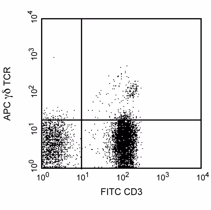
Flow cytometric analysis of TCR γδ expression on human peripheral blood lymphocytes. Human whole blood was costained with FITC Mouse Anti-Human CD3 (Cat. No. 561807/561806/555332/555916) and APC Mouse Anti-Human TCR γδ (Cat. No. 555718/561049). Erythrocytes were lysed with BD FACS™ Lysing Solution (Cat. No. 349202). Two color dot-plots were derived from gated events with the side and forward light-scatter characteristics of viable lymphocytes. Flow cytometric analysis was carried out on a BD FACScan™ System.
Flow cytometric analysis of TCR γδ expression on human peripheral blood lymphocytes. Human whole blood was costained with FITC Mouse Anti-Human CD3 (Cat. No. 561807/561806/555332/555916) and APC Mouse Anti-Human TCR γδ (Cat. No. 555718/561049). Erythrocytes were lysed with BD FACS™ Lysing Solution (Cat. No. 349202). Two color dot-plots were derived from gated events with the side and forward light-scatter characteristics of viable lymphocytes. Flow cytometric analysis was carried out on a BD FACScan™ System.


 全部商品分类


 下载产品说明书
 用小程序,查商品更便捷


 收藏
收藏
 对比
对比 咨询
咨询
