


 全部商品分类


1/3
BD IMag™ Anti-Mouse Ter-119 Particles - DM

品牌: BD Pharmingen
 下载产品说明书
 用小程序,查商品更便捷


 收藏
收藏
 对比
对比 咨询
咨询反应种属:
Mouse (QC Testing)
实验应用:
Cell separation (Routinely Tested)
产品介绍
产品介绍
产品信息
抗原名称
Ly-76 (TER-119)

免疫原
Mouse Fetal Liver
简单描述
The TER-119 antibody specifically binds to a 52 kDa molecule associated with glycophorin A on cells of the erythroid lineage in embryonic yolk sac, fetal liver, newborn liver, adult bone marrow, adult peripheral blood, and adult lymphoid organs. The TER-119 antigen is expressed on erythroid cells from pro-erythroblast through mature erythrocyte stages, but not on cells with BFU-E or CFU-E activities. The TER-119 epitope is not detected on hematopoietic stem cells, lymphoid cells, myeloid cells, or erythroleukemia lines. The TER-119 mAb is a component of the "lineage cocktail" used in studies of hematopoietic progenitors to detect, or deplete cells committed to the hematopoietic lineages.
商品描述
TER-119
The TER-119 antibody specifically binds to a 52 kDa molecule associated with glycophorin A on cells of the erythroid lineage in embryonic yolk sac, fetal liver, newborn liver, adult bone marrow, adult peripheral blood, and adult lymphoid organs. The TER-119 antigen is expressed on erythroid cells from pro-erythroblast through mature erythrocyte stages, but not on cells with BFU-E or CFU-E activities. The TER-119 epitope is not detected on hematopoietic stem cells, lymphoid cells, myeloid cells, or erythroleukemia lines. The TER-119 mAb is a component of the "lineage cocktail" used in studies of hematopoietic progenitors to detect, or deplete cells committed to the hematopoietic lineages.
同种型
Rat WI, also known as Wistar (outbred) IgG2b, κ
克隆号
克隆 TER-119 (RUO)
应用
实验应用
Cell separation (Routinely Tested)
反应种属
Mouse (QC Testing)
目标/特异性
Ly-76 (TER-119)
制备和贮存
存储溶液
Aqueous buffered solution containing BSA and ≤0.09% sodium azide.
数据库链接
Entrez-Gene ID
104231
参考图片
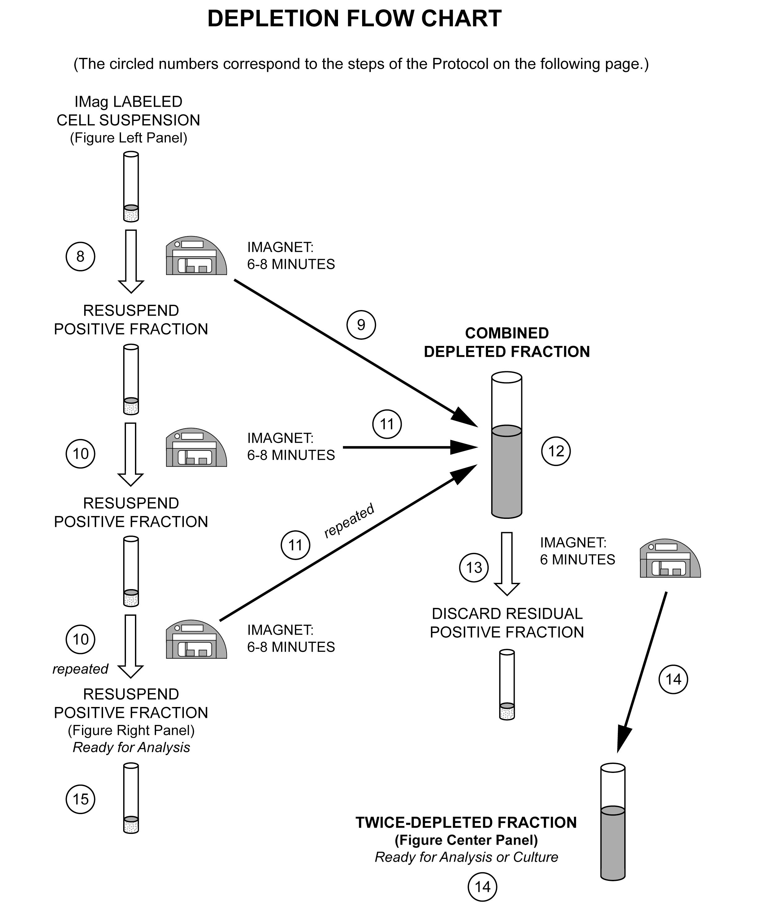
Depletion of mouse TER-119-positive cells from spleen cells. Splenocytes were labeled with BD IMag™ anti-mouseTER-119 particles- DM as described in the protocol. After labeling, the cells were separated using the BD IMagnet™, and the negative (TER-119-) and positive (TER-119+) fractions were collected. Please refer to the Separation Flow Chart to identify the separated cell populations represented in this figure. For flow cytometric analysis, fresh spleen cells (left panel), the depleted fraction (center panel), and the positive fraction (right panel) were stained with PE anti-mouse TER-119 mAb TER-119 (Cat. No. 553673) and FITC anti-mouse CD45 mAb 30-F11 (Cat. No.553080). The percent TER-119+ cells in each sample is given.
声明 :本官网所有报价均为常温或者蓝冰运输价格,如有产品需要干冰运输,需另外加收干冰运输费。