The C2 monoclonal antibody specifically binds to CD71, the transferrin receptor. CD71 is a disulfide-linked homodimer of 95-kDa subunits. CD71 mediates one of the cellular mechanisms for iron uptake, and its expression is regulated according to the cell's iron requirements. It is expressed at high levels on developing erythroid cells, and it is upregulated after mitogenic activation of B or T lymphocytes. The C2 monoclonal antibody selectivity inhibits some types of T- and B-cell activation by down-regulation of transferrin receptor expression, but it does not block binding of transferrin.
This antibody is conjugated to BD Horizon BV605 which is part of the BD Horizon Brilliant™ Violet family of dyes. With an Ex Max of 407-nm and Em Max of 602-nm, BD Horizon BV605 can be excited by a violet laser and detected with a standard 610/20-nm filter set. BD Horizon BV605 is a tandem fluorochrome of BD Horizon BV421 and an acceptor dye with an Em max at 605-nm. Due to the excitation of the acceptor dye by the green (532 nm) and yellow-green (561 nm) lasers, there will be significant spillover into the PE and BD Horizon PE-CF594 detectors off the green or yellow-green lasers. BD Horizon BV605 conjugates are very bright, often exhibiting brightness equivalent to PE conjugates and can be used as a third color off of the violet laser.
For optimal and reproducible results, BD Horizon Brilliant Stain Buffer should be used anytime two or more BD Horizon Brilliant dyes are used in the same experiment. Fluorescent dye interactions may cause staining artifacts which may affect data interpretation. The BD Horizon Brilliant Stain Buffer was designed to minimize these interactions. More information can be found in the Technical Data Sheet of the BD Horizon Brilliant Stain Buffer (Cat. No. 563794).
商品描述
C2
The C2 monoclonal antibody specifically binds to CD71, the transferrin receptor. CD71 is a disulfide-linked homodimer of 95-kDa subunits. CD71 mediates one of the cellular mechanisms for iron uptake, and its expression is regulated according to the cell's iron requirements. It is expressed at high levels on developing erythroid cells, and it is upregulated after mitogenic activation of B or T lymphocytes. The C2 monoclonal antibody selectivity inhibits some types of T- and B-cell activation by down-regulation of transferrin receptor expression, but it does not block binding of transferrin.
This antibody is conjugated to BD Horizon BV605 which is part of the BD Horizon Brilliant™ Violet family of dyes. With an Ex Max of 407-nm and Em Max of 602-nm, BD Horizon BV605 can be excited by a violet laser and detected with a standard 610/20-nm filter set. BD Horizon BV605 is a tandem fluorochrome of BD Horizon BV421 and an acceptor dye with an Em max at 605-nm. Due to the excitation of the acceptor dye by the green (532 nm) and yellow-green (561 nm) lasers, there will be significant spillover into the PE and BD Horizon PE-CF594 detectors off the green or yellow-green lasers. BD Horizon BV605 conjugates are very bright, often exhibiting brightness equivalent to PE conjugates and can be used as a third color off of the violet laser.
For optimal and reproducible results, BD Horizon Brilliant Stain Buffer should be used anytime two or more BD Horizon Brilliant dyes are used in the same experiment. Fluorescent dye interactions may cause staining artifacts which may affect data interpretation. The BD Horizon Brilliant Stain Buffer was designed to minimize these interactions. More information can be found in the Technical Data Sheet of the BD Horizon Brilliant Stain Buffer (Cat. No. 563794).
同种型
Rat WF, also known as Wistar Furth IgG1, κ
克隆号
克隆 C2 (also known as C2F2) (RUO)
浓度
0.2 mg/ml
产品详情
BV605
The BD Horizon Brilliant Violet™ 605 (BV605) dye is part of the BD Horizon Brilliant Violet™ family of dyes. This tandem fluorochrome is comprised of a BV421 donor with an excitation maximum (Ex Max) of 407-nm and an acceptor dye with an emission maximum (Em Max) at 605-nm. BV605, driven by BD innovation, is designed to be excited by the violet laser (405-nm) and detected using an optical filter centered near 610-nm (e.g., a 610/20-nm bandpass filter). The acceptor dye can be excited by the yellow-green (561-nm) laser resulting in cross-laser excitation and fluorescence spillover. Please ensure that your instrument’s configurations (lasers and optical filters) are appropriate for this dye.
Aqueous buffered solution containing BSA and ≤0.09% sodium azide.
文献
文献
研发参考(5)
1. Fujimoto T. GPI-anchored proteins, glycosphingolipids, and sphingomyelin are sequestered to caveolae only after crosslinking. J Histochem Cytochem. 1996; 44(8):929-941. (Clone-specific: Immunofluorescence).
2. Kemp JD, Thorson JA, Gomez F, Smith KM, Cowdery JS, Ballas ZK. Inhibition of lymphocyte activation with anti-transferrin receptor Mabs: a comparison of three reagents and further studies of their range of effects and mechanism of action. Cell Immunol. 1989; 122(1):218-230. (Clone-specific: Activation, Inhibition).
3. Kemp JD, Thorson JA, McAlmont TH, Horowitz M, Cowdery JS, Ballas ZK. Role of the transferrin receptor in lymphocyte growth: a rat IgG monoclonal antibody against the murine transferrin receptor produces highly selective inhibition of T and B cell activation protocols. J Immunol. 1987; 138(8):2422-2426. (Immunogen: Activation, Immunoprecipitation, Inhibition).
4. Lok CN, Loh TT. Regulation of transferrin function and expression: review and update. Biol Signals Recept. 1998; 7(3):157-178. (Biology).
5. Thorson JA, Smith KM, Gomez F, Naumann PW, Kemp JD. Role of iron in T cell activation: TH1 clones differ from TH2 clones in their sensitivity to inhibition of DNA synthesis caused by IgG Mabs against the transferrin receptor and the iron chelator deferoxamine. Cell Immunol. 1991; 134(1):126-137. (Biology).
参考图片
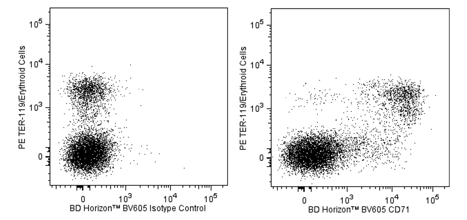
Two-color flow cytometric analysis of CD71 expression on developing mouse erythroid cells. BALB/c mouse bone marrow cells were stained with PE Rat Anti-Mouse TER-119/Erythroid Cells antibody (Cat. No. 553673/561071) and either BD Horizon™ BV605 Rat IgG1, κ Isotype Control (Cat. No. 562993, Left Panel) or BD Horizon™ BV605 Rat Anti-Mouse CD71 (Cat. No. 563013, Right Panel). The two-color flow cytometric dot plots show the correlated expression of CD71 (or Ig Isotype control staining) versus TER-119 for gated events with the forward and side light-scatter characteristics of viable bone marrow cells. Flow cytometry was performed using a BD™ LSR II Flow Cytometer System
Two-color flow cytometric analysis of CD71 expression on developing mouse erythroid cells. BALB/c mouse bone marrow cells were stained with PE Rat Anti-Mouse TER-119/Erythroid Cells antibody (Cat. No. 553673/561071) and either BD Horizon™ BV605 Rat IgG1, κ Isotype Control (Cat. No. 562993, Left Panel) or BD Horizon™ BV605 Rat Anti-Mouse CD71 (Cat. No. 563013, Right Panel). The two-color flow cytometric dot plots show the correlated expression of CD71 (or Ig Isotype control staining) versus TER-119 for gated events with the forward and side light-scatter characteristics of viable bone marrow cells. Flow cytometry was performed using a BD™ LSR II Flow Cytometer System


 全部商品分类


 下载产品说明书
 用小程序,查商品更便捷


 收藏
收藏
 对比
对比 咨询
咨询
