


 全部商品分类


TGF-b1,2,3 MAb (Cl 1 (25 ug)


 下载产品说明书 下载SDS
 用小程序,查商品更便捷


 收藏
收藏
 对比
对比 咨询
咨询
Scientific Data
 View Larger
TGF‑ beta 1 Inhibition of IL‑4-dependent Cell Proliferation and Neutralization by TGF‑ beta 1, 2, 3 Antibody. Recombinant Human TGF-beta 1 (Catalog # 240-B) inhibits Recombinant Mouse IL-4 (Catalog # 404-ML) induced proliferation in the HT-2 mouse T cell line in a dose-dependent manner (orange line). Inhibition of Recombinant Mouse IL-4 (7.5 ng/mL) activity elicited by Recombinant Human TGF-beta 1 (1 ng/mL) is neutralized (green line) by increasing concentrations of Mouse Anti-TGF-beta 1, 2, 3 Monoclonal Antibody (Catalog # MAB1835). The ND50 is typically 0.25-1.25 µg/mL.
 View Larger
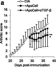
Detection of Mouse TGF beta 1/2/3 by In vivo assay Apoptotic cell-induced immunomodulation in mice with collagen-induced arthritis (CIA) is dependent on transforming growth factor (TGF)-beta. CIA mice that received or did not receive apoptotic cells, with or without anti-TGF-beta blocking antibody were scored daily (a). Data are shown as mean ± SEM of five mice per group from one of two representative experiments; *p < 0.05, **p < 0.01 (Friedman test analysis of variance (ANOVA) with Dunn's multiple comparisons test). Anti-collagen IgG2a antibodies (Ab) were quantified in plasma (b). Data from 5 to 16 mice from two to three independent experiments (no difference, Kruskal-Wallis test ANOVA with Dunn’s multiple comparisons test). Lymph node cells were collected, cultured, and T cell proliferation in response to collagen protein or CD3-specific Ab stimulations was evaluated (c). Data are shown as mean ± SEM from one of two representative experiments; *p < 0.05, **p < 0.01 (nonparametric unpaired t test). Percent and absolute number of Foxp3+ T regulatory cells (Treg) were evaluated in the spleen (d) and suppressive assays were performed by isolating and adding Treg at a different ratio into collagen-specific cultures and responder T cell proliferation was assessed (e). Data are shown as mean ± SEM from one experiment (d Kruskal-Wallis test ANOVA with Dunn’s multiple comparisons test; e Friedman test ANOVA with Dunn’s multiple comparisons test). Plasmacytoid dendritic cells (pDC), conventional dendritic cells (cDC) and macrophages (macro) were isolated and cultured with naïve allogenic T cells to determine Treg polarization (f), and their response to TLR ligand was assessed through evaluation of CD40 costimulatory molecule expression (g). Data from individual mouse plus mean (black bar) from one experiment representative of four are shown; *p < 0.05, ***p < 0.001 (f ordinary one-way ANOVA with Tukey’s multiple comparisons test; g Kruskal-Wallis test ANOVA with Dunn’s multiple comparisons test). BrDU 5-Bromo-2’-deoxyuridine Image collected and cropped by CiteAb from the following publication (https://arthritis-research.biomedcentral.com/articles/10.1186/s13075-016-1084-0), licensed under a CC-BY license. Not internally tested by R&D Systems.
 View Larger
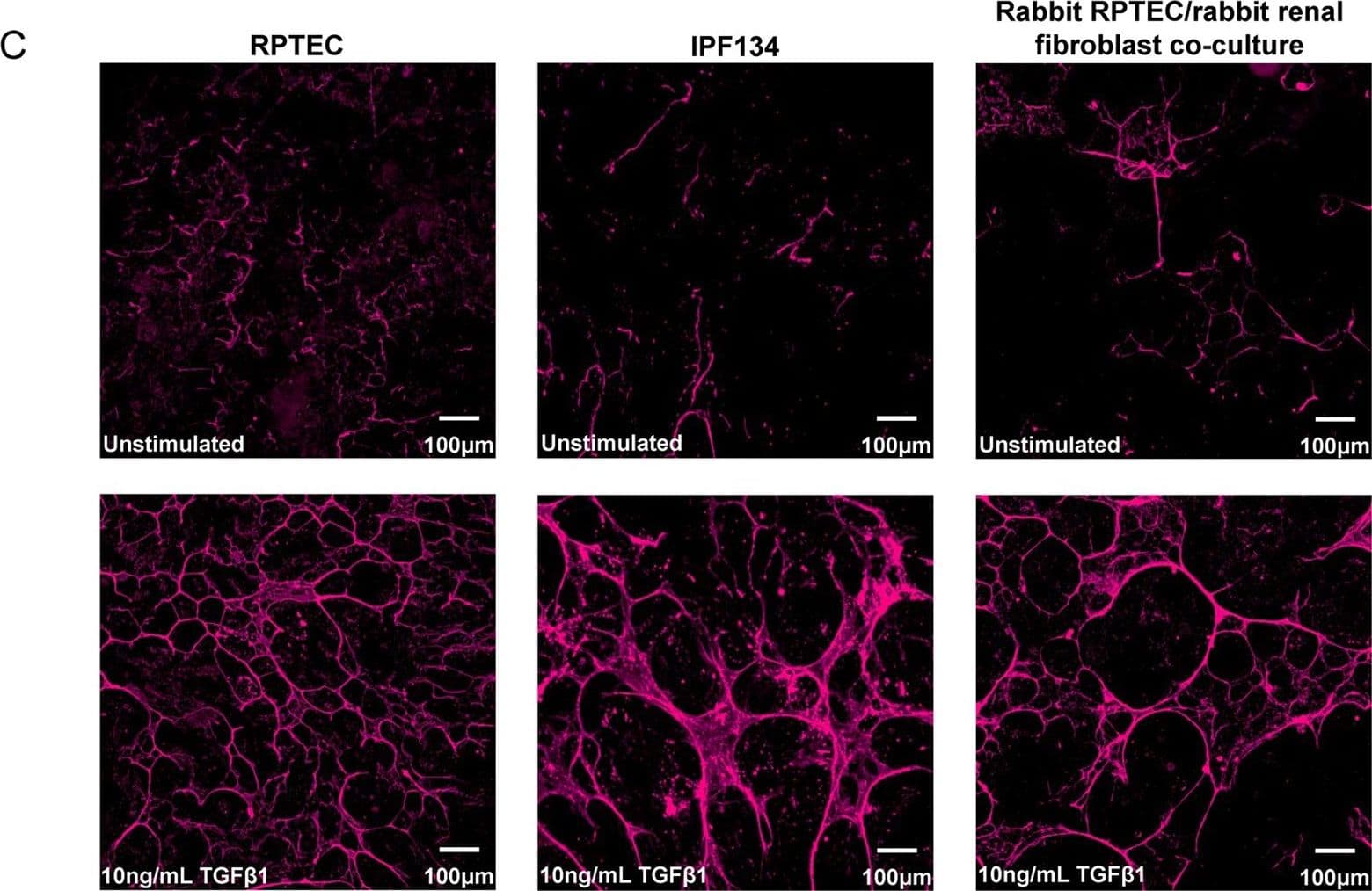
Detection of Human TGF beta 1/2/3 by Immunocytochemistry/Immunofluorescence TGF beta 1 stimulates the expression of ECM components in human and rabbit primary cells. (A) Selected ECM mRNA transcripts were measured using qRT-PCR in human RPTEC or human IPF134 following 48 hr stimulation with TGF beta 1 (10 ng/mL). Data show transcript abundance relative to the unstimulated control (indicated by the dashed line) and are shown as mean ± SD of four (RPTEC) or seven (IPF134) independent experiments. ****P < 0.0001; ****P < 0.001; **P < 0.01; Student’s t-test. (B) The accumulation of ECM in human RPTEC (i) or human IPF (ii) cells stimulated with 10ng/mL TGF beta 1 was evaluated via measurement of the incorporation of 14C-labelled amino acids into the deposited ECM. Data presented are the mean ± SD of three independent experiments. **P < 0.01; Student’s t-test. (C) Increased mature ECM accumulation following treatment of cells with a pro-fibrotic stimulus can be visualised following in-situ fluorescent staining using the Flamingo dye. Cells from three different systems (primary human RPTEC; primary human IPF134; co-culture of primary rabbit RPTEC with primary rabbit renal fibroblasts) were cultured in the absence (top) or presence (bottom) of 10 ng/ml TGF beta 1 before decellularised matrix was fixed and stained in situ with Flamingo fluorescent dye. Images show a single field. Image collected and cropped by CiteAb from the following publication (https://pubmed.ncbi.nlm.nih.gov/28855577), licensed under a CC-BY license. Not internally tested by R&D Systems.
TGF-beta 1, 2, 3 Antibody Summary
Applications
Human TGF-beta 1 Sandwich Immunoassay
Please Note: Optimal dilutions should be determined by each laboratory for each application. General Protocols are available in the Technical Information section on our website.
Background: TGF-beta 1, 2, 3
TGF-beta 1, -2, and -3 are a closely related group of proteins (70‑80% sequence homology) that are produced by many cell types and function as growth and differentiation factors. The active forms of TGF-beta 1, -2, and -3 are disulfide-linked homodimers.
- Ayala A. et al. (1992) FASEB J. 6:A1604.
- Roberts A.B. and Sporn M.B., eds. (1990) Peptide Growth Factors and Their Receptors I, Springer-Verlag, 419.
- Dasch J.R. et al. (1989) J. Immunol. 142:1536.
Preparation and Storage
- 12 months from date of receipt, -20 to -70 °C as supplied.
- 1 month, 2 to 8 °C under sterile conditions after reconstitution.
- 6 months, -20 to -70 °C under sterile conditions after reconstitution.
参考图片
TGF‑ beta in PC-3 Human Cell Line. TGF‑ beta 1, 2, 3 was detected in immersion fixed PC-3 human prostate cancer cell line using 10 µg/mL Mouse Anti-TGF‑ beta 1, 2, 3 Monoclonal Antibody (Catalog # MAB1835) for 3 hours at room temperature. Cells were stained with the NorthernLights™ 557-conjugated Anti-Mouse IgG Secondary Antibody (red; Catalog # NL007) and counterstained with DAPI (blue). View our protocol for Fluorescent ICC Staining of Cells on Coverslips.
TGF-beta in Human Skin. TGF-beta 1, 2, 3 was detected in immersion fixed paraffin-embedded human skin using 25 µg/mL Mouse
Anti-TGF-beta 1, 2, 3 Monoclonal Antibody (Catalog # MAB1835) overnight at 4 °C. Tissues was stained using the Anti-Mouse HRP-DAB Cell and Tissue Staining Kit (brown; Catalog # CTS002) and counterstained with Haematoxylin (blue). View our protocol for Chromogenic IHC Staining of Paraffin-embedded Tissue Sections.
TGF‑ beta 1 Inhibition of IL‑4-dependent Cell Proliferation and Neutralization by TGF‑ beta 1, 2, 3 Antibody. Recombinant Human TGF‑ beta 1 (Catalog # 240‑B) inhibits Recombinant Mouse IL‑4 (Catalog # 404-ML) induced proliferation in the HT‑2 mouse T cell line in a dose-dependent manner (orange line). Inhibition of Recombinant Mouse IL‑4 (7.5 ng/mL) activity elicited by Recombinant Human TGF‑ beta 1 (1 ng/mL) is neutralized (green line) by increasing concentrations of Mouse Anti-TGF‑ beta 1, 2, 3 Monoclonal Antibody (Catalog # MAB1835). The ND50 is typically
0.25‑1.25 µg/mL.