


1/44

品牌: CST
 下载产品说明书
 用小程序,查商品更便捷


 收藏
收藏
 对比
对比 咨询
咨询
产品介绍
产品信息
抗原名称
TGF-beta Fibrosis Pathway

来源纯化
Monoclonal antibody is produced by immunizing animals with a synthetic peptide corresponding to residues near the amino terminus of human α-Smooth Muscle Actin protein. Monoclonal antibody is produced by immunizing animals with a synthetic peptide corresponding to residues surrounding Phe1197 of human COL1A1 protein. Monoclonal antibody is produced by immunizing animals with a synthetic peptide corresponding to residues surrounding His198 of human Smad2/3 protein. Monoclonal antibody is produced by immunizing animals with a synthetic peptide corresponding to residues near the amino terminus of mouse Smad2 protein. Monoclonal antibody is produced by immunizing animals with a synthetic peptide corresponding to residues surrounding Ser465/467 of human Smad2 protein. Monoclonal antibody is produced by immunizing animals with a synthetic peptide corresponding to residues near the amino terminus of human YKL-40 protein. Monoclonal antibody is produced by immunizing animals with a synthetic peptide corresponding to residues surrounding Ser465/467 of human Smad2 protein. Monoclonal antibody is produced by immunizing animals with a synthetic peptide corresponding to a region in the carboxy terminus of TGF-β1 protein. Monoclonal antibody is produced by immunizing animals with recombinant protein specific to the amino terminus of human TGF-β Receptor II protein.
简单描述
Antibody Sampler Kit for studying YKL-40/Smad3 (Ser423/Ser425) phosphate/Smad3/COL1A1/Smad2/Smad2 (Ser465/Ser467) phosphate/ACTA2 (alpha actin, smooth muscle)/TGFB1/TGFBR2 in the research area.
研究领域
癌症,细胞生物学,发育生物学与干细胞研究,纤维化,神经科学
应用
目标/特异性
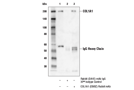
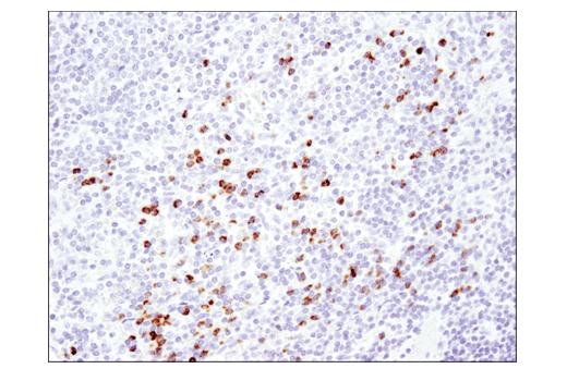
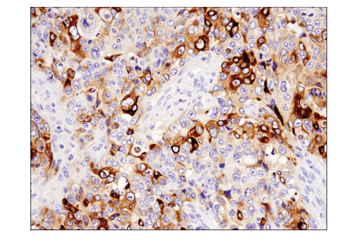
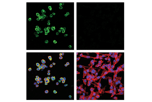
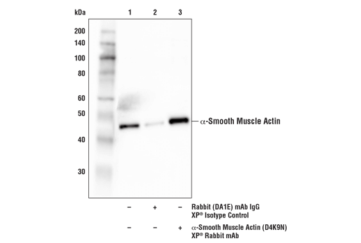
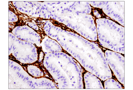
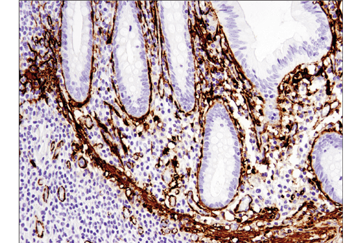
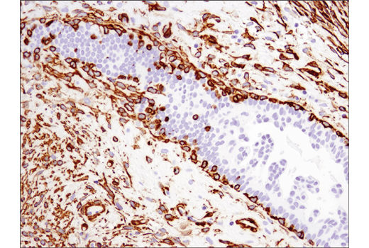
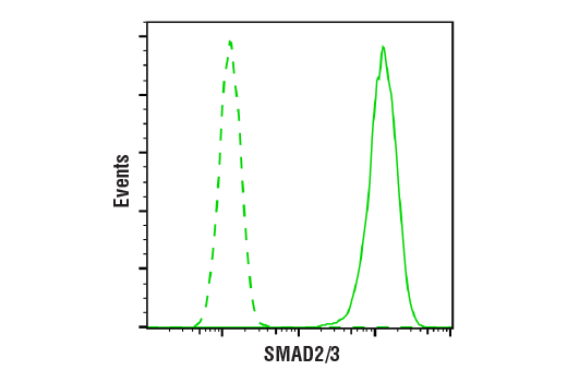
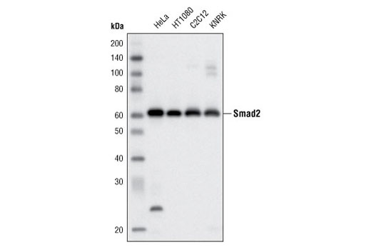
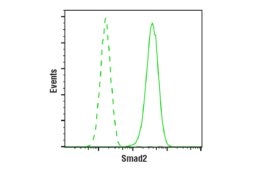
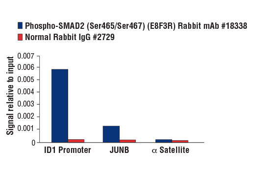
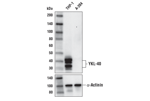
Specificity/Sensitivity
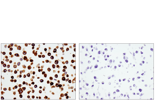
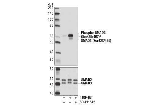
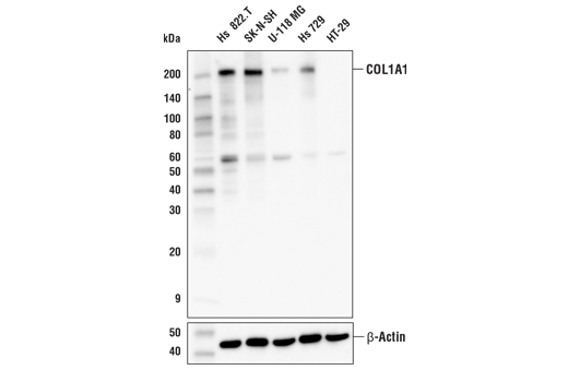
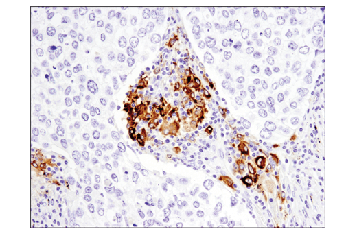
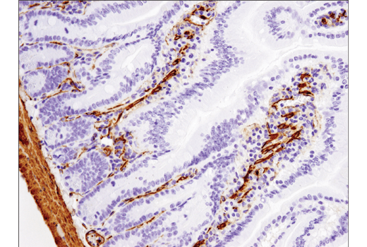
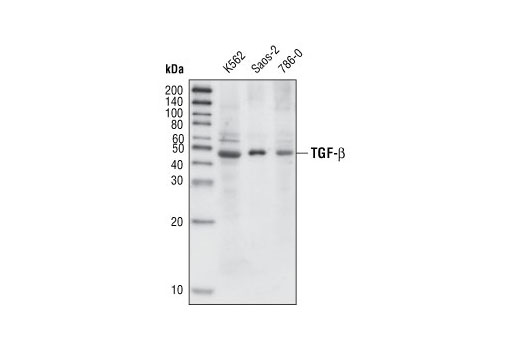
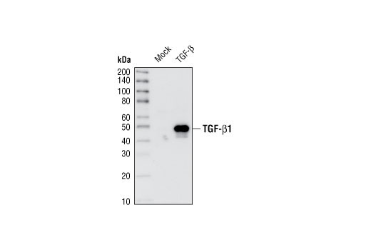
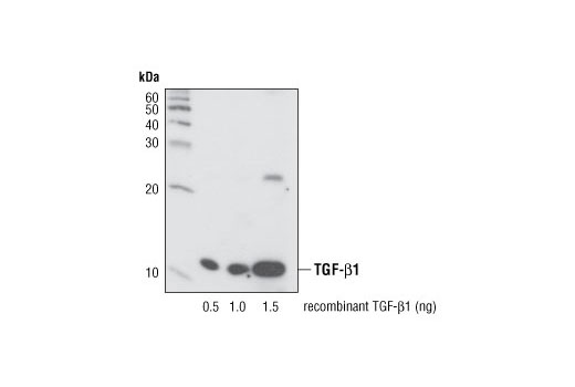
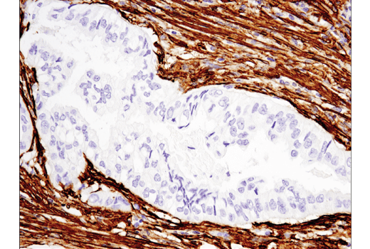
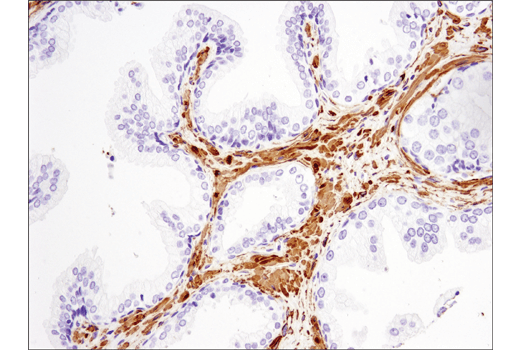
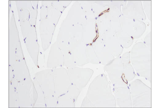
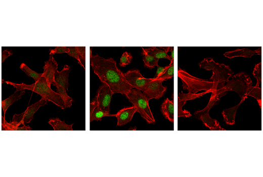
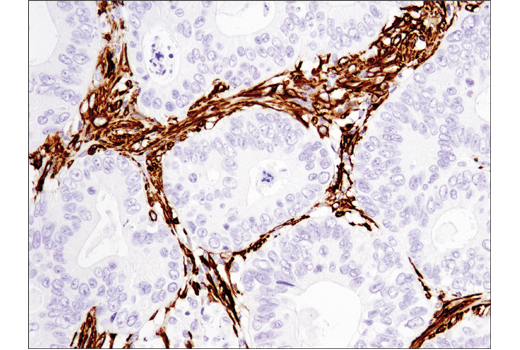
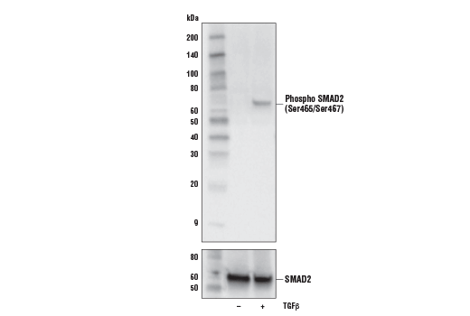
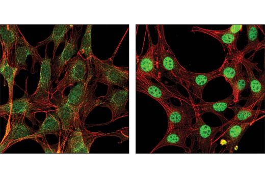
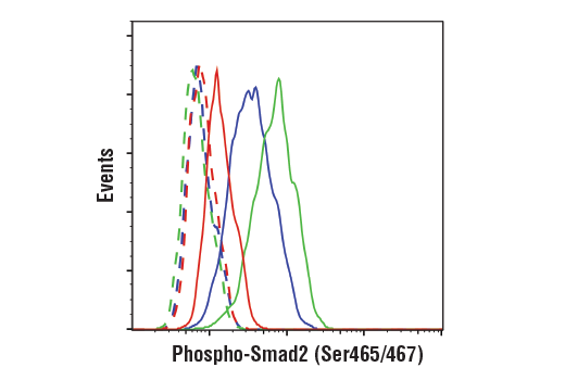
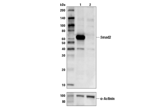
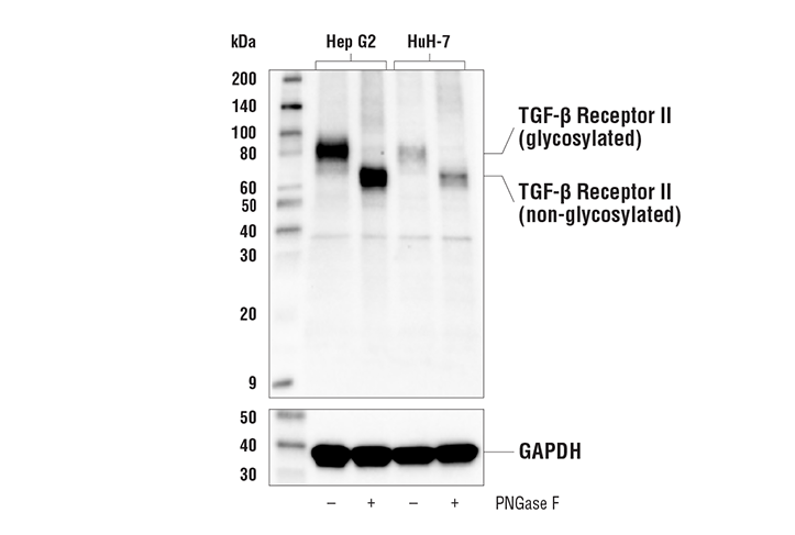
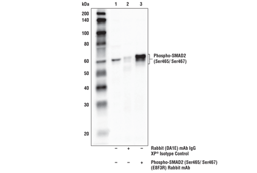
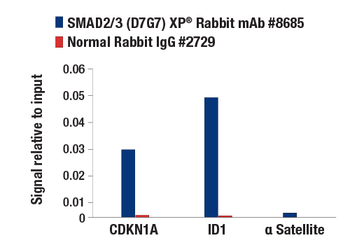
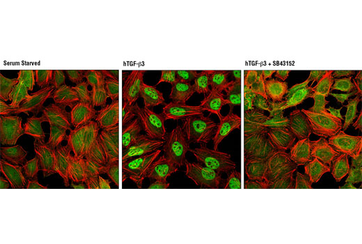
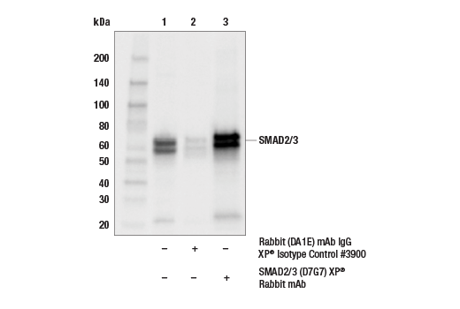
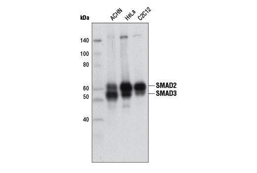
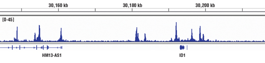
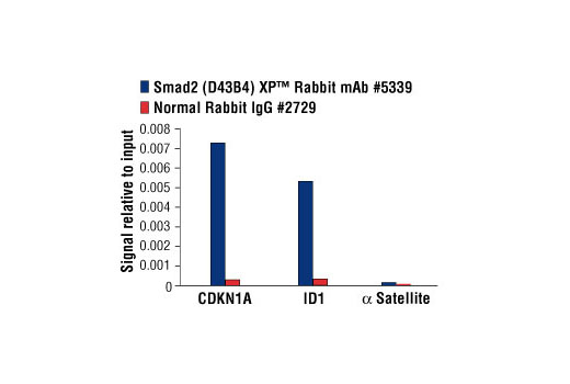
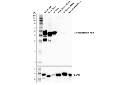
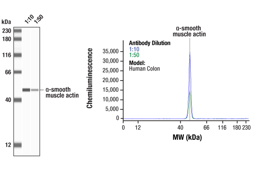
α-Smooth Muscle Actin (D4K9N) XP® Rabbit mAb recognizes endogenous levels of total α-smooth muscle protein. COL1A1 (E8I9Z) Rabbit mAb recognizes endogenous levels of total COL1A1 protein. Smad2/3 (D7G7) XP® Rabbit mAb recognizes endogenous levels of total Smad2/3 protein. Smad2 (D43B4) XP® Rabbit mAb detects endogenous levels of total Smad2 protein. This antibody does not cross-react with Smad3. Phospho-Smad2 (Ser465/Ser467) (E8F3R) Rabbit mAb recognizes endogenous levels of Smad2 protein when phosphorylated at Ser465 and Ser467. YKL-40 (E2L1M) Rabbit mAb recognizes endogenous levels of total YKL-40 protein. Phospho-Smad2 (Ser465/467)/Smad3 (Ser423/425) (D27F4) Rabbit mAb recognizes endogenous levels of Smad2 protein when phosphorylated at Ser465 and Ser467. This antibody also recognizes endogenous levels of Smad3 protein when phosphorylated at Ser422 only or at both Ser423 and Ser425. TGF-β (56E4) Rabbit mAb detects recombinant TGF-β1 and TGF-β3 proteins. The antibody also detects endogenous levels of the TGF-β precursor proteins. TGF-β Receptor II (E5M6F) Rabbit mAb recognizes endogenous levels of total TGF-β Receptor II protein. This antibody does not cross-react with TGF-β Receptor I protein.
背景
背景
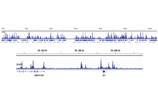
Transforming growth factor-β (TGF-β) superfamily members are critical regulators of cell proliferation and differentiation, developmental patterning and morphogenesis, and disease pathogenesis (1-4). In the context of fibrosis, TGF-β signaling to SMAD2/3 is one of the biggest drivers of the profibrotic program (5).TGF-β elicits signaling through three cell surface receptors: type I (RI), type II (RII), and type III (RIII). In response to ligand binding, the type II receptors form stable heterotrimeric complexes with the type I receptors, allowing phosphorylation and activation of type I receptor kinase. Activated type I receptors associate with SMAD2/3 and phosphorylate them on a conserved carboxy terminal SSXS motif. The phosphorylated SMADs dissociate from the receptor and form a heterotrimeric complex with the co-Smad (Smad4), allowing translocation of the complex to the nucleus. Once in the nucleus, phosphorylated SMAD2/3 targets a subset of DNA binding proteins to regulate the transcriptional program (6-8).In the context of fibrosis, SMAD2/3 activation upregulates expression of profibrotic genes such as COL1A1 and other ECM modulators that modify the extracellular matrix of the tissue. (9). TGF-β/ SMAD2/3 signaling also induces expression of α-Smooth Muscle Actin in fibroblasts, causing transformation of these cells to myofibroblasts (10). Myofibroblasts further modify the ECM, causing excessive accumulation of collagens and other ECM components. Injury to the tissue attracts macrophages and other immune cells and the fibrotic tissue soon becomes a site of inflammation (11). In this pro-fibrotic, pro-inflammatory environment, YKL-40, also known as Chitinase-3-like protein 1 (CHI3L1), is secreted. YKL-40 is a pro-inflammatory glycoprotein that also contributes to the progression of fibrosis (12). Measurement of collagen content, α-Smooth Muscle Actin, and the release of YKL-40 are predictive of fibrotic activity.
1.Massagué, J. et al. (2000) Cell 103, 295-309.
2.de Caestecker, M.P. et al. (2000) J Natl Cancer Inst 92, 1388-402.
3.Derynck, R. et al. (2001) Nat Genet 29, 117-29.
4.Miyazono, K. et al. (2000) Adv Immunol 75, 115-57.
5.Meng, X.M. et al. (2016) Nat Rev Nephrol 12, 325-38.
6.Wu, G. et al. (2000) Science 287, 92-7.
7.Attisano, L. and Wrana, J.L. (2002) Science 296, 1646-7.
8.Moustakas, A. et al. (2001) J Cell Sci 114, 4359-69.
9.Bagalad, B.S. et al. J Oral Maxillofac Pathol 21, 462-3.
10.Mack, M. (2018) Matrix Biol 68-69, 106-21.
11.Johansen, J.S. (2006) Dan Med Bull 53, 172-209.
研究领域
癌症,细胞生物学,发育生物学与干细胞研究,纤维化,神经科学
数据库链接
Entrez-Gene ID
1116,4088,1277,4087,59,7040,7048
UniProt ID
P36222,P84022,P02452,Q15796,P62736,P01137,P37173
声明 :本官网所有报价均为常温或者蓝冰运输价格,如有产品需要干冰运输,需另外加收干冰运输费。

 危险品化学品经营许可证(不带存储) 许可证编号:沪(杨)应急管危经许[2022]202944(QY)
  营业执照(三证合一)