


 全部商品分类
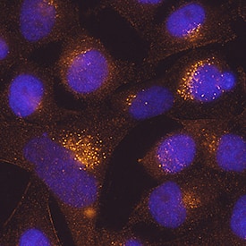


 下载产品说明书 下载SDS
 用小程序,查商品更便捷


 收藏
收藏
 对比
对比 咨询
咨询
Met1-Asn220
Accession # Q9BXS4
Scientific Data
.") View Larger
View LargerTMEM59 in HeLa Human Cell Line. TMEM59 was detected in immersion fixed HeLa human cervical epithelial carcinoma cell line using Mouse Anti-Human TMEM59 Monoclonal Antibody (Catalog # MAB9118) at 25 µg/mL for 3 hours at room temperature. Cells were stained using the NorthernLights™ 557-conjugated Anti-Mouse IgG Secondary Antibody (red; Catalog # NL007) and counterstained with DAPI (blue). Specific staining was localized to cytoplasm. View our protocol for Fluorescent ICC Staining of Cells on Coverslips.
Human TMEM59 Antibody Summary
Met1-Asn220
Accession # Q9BXS4
Applications
Please Note: Optimal dilutions should be determined by each laboratory for each application. General Protocols are available in the Technical Information section on our website.
Background: TMEM59
TMEM59 is a 324 aa protein with a 21 amino acid transmembrane domain which localizes to the Golgi compartment where it modulates the O-glycosylation and complex N-glycosylation maturation steps of several proteins including APP, BACE1, SEAP and PRNP. It may retain APP in the Golgi and inhibits amyloid beta generation as well APP cleavage by alpha and beta secretases. TMEM59 also contains a 19 aa peptide that acts as a regulator of autophagy in response to bacterial infection by promoting LC3 activation through interaction with ATG16L1, targeting its own endosomal compartment to lysosomes in response to aggregation during S.areus infection.
- Ullrich S, et al, J Biol Chem. 2010 Jul 2;285(27):20664
- Boada-Romero E, et al, EMBO J. 2013 Feb 20;32(4):566
Preparation and Storage
- 12 months from date of receipt, -20 to -70 °C as supplied.
- 1 month, 2 to 8 °C under sterile conditions after reconstitution.
- 6 months, -20 to -70 °C under sterile conditions after reconstitution.
参考图片
TMEM59 in HeLa Human Cell Line. TMEM59 was detected in immersion fixed HeLa human cervical epithelial carcinoma cell line using Mouse Anti-Human TMEM59 Monoclonal Antibody (Catalog # MAB9118) at 25 µg/mL for 3 hours at room temperature. Cells were stained using the NorthernLights™ 557-conjugated Anti-Mouse IgG Secondary Antibody (red; Catalog # NL007) and counterstained with DAPI (blue). Specific staining was localized to cytoplasm. View our protocol for Fluorescent ICC Staining of Cells on Coverslips.