


 全部商品分类
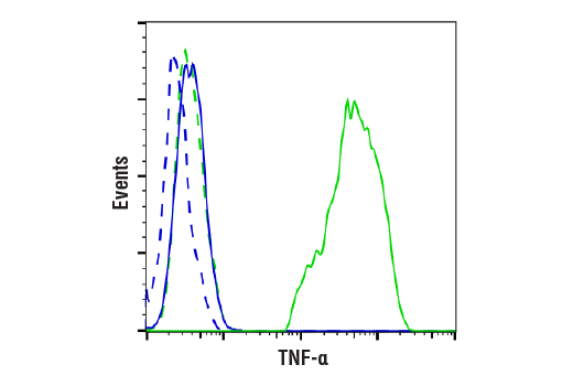


 用小程序,查商品更便捷


Monoclonal antibody is produced by immunizing animals with a recombinant mouse TNF-α protein.
Product Usage Information
| Application | Dilution |
|---|---|
| Western Blotting | 1:1000 |
| Immunoprecipitation | 1:50 |
| Immunofluorescence (Immunocytochemistry) | 1:100 - 1:400 |
| Flow Cytometry (Fixed/Permeabilized) | 1:100 - 1:400 |
Specificity/Sensitivity
Species Reactivity:
Mouse
Supplied in 10 mM sodium HEPES (pH 7.5), 150 mM NaCl, 100 µg/ml BSA, 50% glycerol and less than 0.02% sodium azide. Store at –20°C. Do not aliquot the antibody.
For a carrier free (BSA and azide free) version of this product see product #57485.
参考图片
Flow cytometric analysis of Raw 264.7 cells, untreated (blue) or treated with LPS (100 ng/ml, 6 hr) and Brefeldin A #9972 (300 ng/ml, last 3 hr of treatment) (green), using TNF-α (D2D4) XP® Rabbit mAb (solid lines) or concentration-matched Rabbit (DA1E) mAb IgG XP® Isotype Control #3900 (dashed lines). Anti-rabbit IgG (H+L), F(ab')2 Fragment (Alexa Fluor® 488 Conjugate) #4412 was used as a secondary antibody.
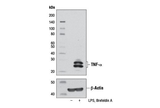
Western blot analysis of extracts from Raw 264.7 cells, untreated (-) or treated (+) with LPS (100 ng/mL, 6 hr) and Brefeldin A #9972 (300 ng/mL, last 3 hr of stimulation), using TNF-α (D2D4) XP® Rabbit mAb (upper) or β-Actin (D6A8) Rabbit mAb #8457 (lower).
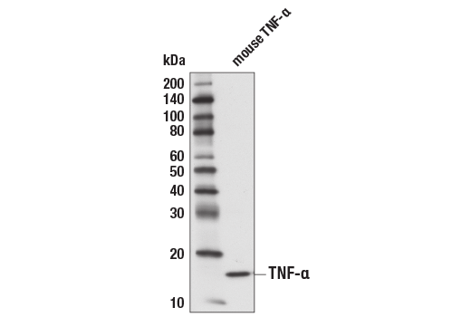
Western blot analysis of 1 ng recombinant mouse TNF-α #5178 using TNF-α (D2D4) XP® Rabbit mAb.
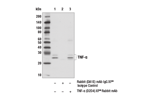
Immunoprecipitation of TNF-α from Raw 264.7 cell extracts, treated with LPS (100 ng/mL, 6 hr) and Brefeldin A #9972 (300 ng/mL, last 3 hr of stimulation), using Rabbit (DA1E) mAb IgG XP® Isotype Control #3900 (lane 2) or TNF-α (D2D4) XP® Rabbit mAb (lane 3). Lane 1 is 10% input. Western blot analysis was performed using TNF-α (D2D4) XP® Rabbit mAb. Mouse Anti-rabbit IgG (Conformation Specific) (L27A9) #3678 and Anti-mouse IgG, HRP-linked Antibody #7076 were used as sencondary antibodies.
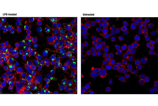
Confocal immunofluorescent analysis of Raw 264.7 cells, treated with LPS (100 ng/mL, 6 hr; left) or untreated (right), using TNF-α (D2D4) XP® Rabbit mAb (green). Actin filaments were labeled with DY-554 phalloidin (red). Blue pseudocolor = DRAQ5® #4084 (fluorescent DNA dye).