


 全部商品分类
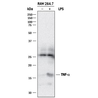


mTNF-a Aff Pur PAb (100 ug)

 下载产品说明书 下载SDS
 用小程序,查商品更便捷


 收藏
收藏
 对比
对比 咨询
咨询
Leu80-Leu235
Accession # P06804
Scientific Data
 View Larger
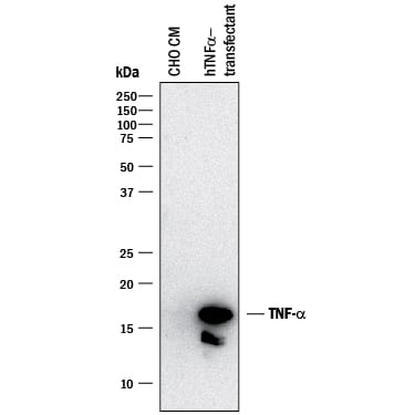
Detection of Human TNF‑ alpha by Western Blot. Western blot shows conditioned media from CHO Chinese hamster ovary cell line either mock transfected or transfected with human TNF- alpha. PVDF membrane was probed with 2 µg/mL of Goat Anti-Human/Mouse TNF-a Antigen Affinity-purified Polyclonal Antibody (Catalog # AF-410-NA) followed by HRP-conjugated Anti-Goat IgG Secondary Antibody (HAF017). Specific bands were detected for TNF-a at approximately 14 and 17 kDa (as indicated). This experiment was conducted under reducing conditions and using Immunoblot Buffer Group 1.
 View Larger
Detection of Mouse TNF‑ alpha by Western Blot. Western blot shows lysates of RAW 264.7 mouse monocyte/macrophage cell line untreated (-) or treated (+) with 10 µg/mL LPS for 4 hours. PVDF membrane was probed with 2 µg/mL of Goat Anti-Human/Mouse TNF-a Antigen Affinity-purified Polyclonal Antibody (Catalog # AF-410-NA) followed by HRP-conjugated Anti-Goat IgG Secondary Antibody (HAF017). Specific bands were detected for TNF-a at approximately 14 and 17 kDa (as indicated). This experiment was conducted under reducing conditions and using Immunoblot Buffer Group 1.
.7 Mouse Cell Line by Immunocytochemistry (ICC).") View Larger
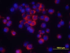
View LargerTNF‑ alpha in RAW 264.7 Mouse Cell Line. TNF-a was detected in immersion fixed RAW 264.7 mouse monocyte/ macrophage cell line treated with LPS using Human/Mouse TNF-a Antigen Affinity-purified Polyclonal Antibody (Catalog # AF-410-NA) at 10 µg/mL for 3 hours at room temperature. Cells were stained using the NorthernLights™ 557-conjugated Anti-Goat IgG Secondary Antibody (red; NL001) and counterstained with DAPI (blue). View our protocol for Fluorescent ICC Staining of Cells on Coverslips.
.") View Larger
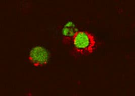
View LargerTNF‑ alpha in Mouse T Cells. TNF‑ alpha was detected in immersion fixed activated mouse T Cells using 15 µg/mL Human/Mouse TNF‑ alpha Antigen Affinity-purified Polyclonal Antibody (Catalog # AF‑410‑NA) for 3 hours at room temperature. Cells were stained (red) and counterstained (green). View our protocol for Fluorescent ICC Staining of Non-adherent Cells.
.") View Larger
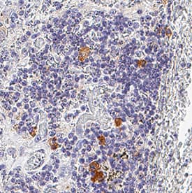
View LargerTNF-alpha in Mouse Spleen Tissue. TNF‑ alpha was detected in immersion fixed paraffin-embedded sections of mouse spleen tissue using Goat Anti-Human/Mouse TNF‑ alpha Antigen Affinity-purified Polyclonal Antibody (Catalog # AF-410-NA) at 3 µg/mL for 1 hour at room temperature followed by incubation with the Anti-Goat IgG VisUCyte™ HRP Polymer Antibody (VC004). Tissue was stained using DAB (brown) and counterstained with hematoxylin (blue). Specific staining was localized to lymphocytes. Staining was performed using our IHC Staining with VisUCyte HRP Polymer Detection Reagents.
 View Larger
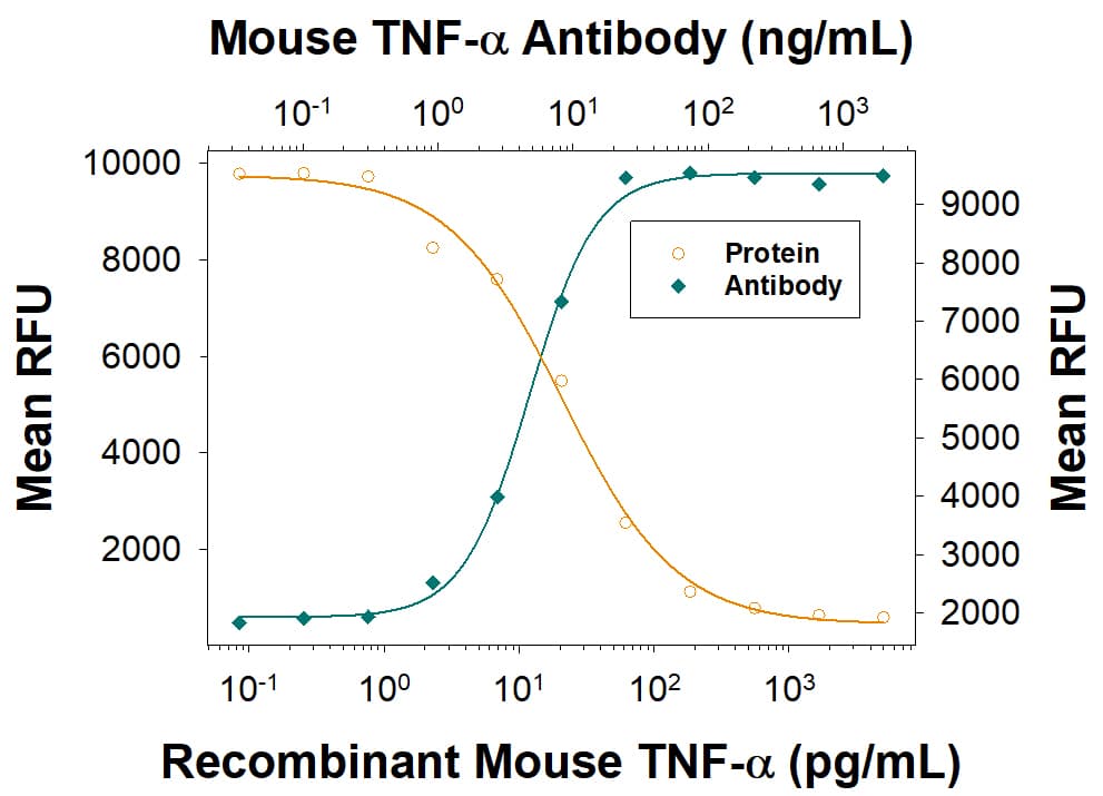
Cytotoxicity Induced by TNF‑ alpha and Neutralization by Mouse TNF‑ alpha Antibody. Recombinant Mouse TNF-a (410-MT) induces cytotoxicity in the the L-929 mouse fibroblast cell line in a dose-dependent manner (orange line). Cytotoxicity elicited by Recombinant Mouse TNF-a (0.1 ng/mL) is neutralized (green line) by increasing concentrations of Human/Mouse TNF-a Antigen Affinity-purified Polyclonal Antibody (Catalog # AF-410-NA). The ND50 is typically 1.5-10 ng/mL in the presence of the metabolic inhibitor actinomycin D.
 View Larger
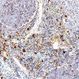
Detection of TNF‑ alpha in Human Spleen. TNF‑ alpha was detected in immersion fixed paraffin-embedded sections of Human Spleen using Goat Anti-Human/Mouse TNF‑ alpha Antigen Affinity-purified Polyclonal Antibody (Catalog # AF-410-NA) at 5 µg/mL for 1 hour at room temperature followed by incubation with the Anti-Goat IgG VisUCyte™ HRP Polymer Antibody (Catalog # VC004). Before incubation with the primary antibody, tissue was subjected to heat-induced epitope retrieval using VisUCyte Antigen Retrieval Reagent-Basic (Catalog # VCTS021). Tissue was stained using DAB (brown) and counterstained with hematoxylin (blue). Specific staining was localized to cytoplasm in lymphocytes. View our protocol for IHC Staining with VisUCyte HRP Polymer Detection Reagents.
 View Larger
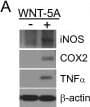
Detection of Mouse TNF-alpha by Western Blot WNT-5A induces a proinflammatory transformation in mouse microglia. (A, B) iNOS, COX-2 and TNF alpha were detected by immunoblotting in lysates from mouse primary microglia after WNT-5A stimulation (ctrl, 300 ng/ml, 6 hours). (B’) shows TNF alpha levels in the supernatant of primary microglia under ctrl conditions and upon WNT-5A stimulation (ctrl, 300 ng/ml, 24 hours; n = 4). At least three experiments are summarized in the bar graphs. Data are normalized to ctrl. Error bars give s.e.m. (C) Microglial proliferation was assessed by an MTT assay monitoring mitochondrial activity, which is proportional to cell number [see Additional file 1: Figure S5]. Stimulation with WNT-5A (300 ng/ml, 24 hours) increased MTT, which was blocked by PTX (100 ng/ml, overnight) or the MEK1/2 (10 μM) inhibitor, SL327. (D). Experiments were done in triplicate and data from three independent experiments are shown. *, P < 0.01; ***, P < 0.001: Error bars show s.e.m.. (E) Cell tracker (red)-stained primary microglia were seeded on top of a collagen matrix in 35 mm glass bottom dishes. One day after ctrl or 300 ng/ml WNT-5A stimulation, invasion was observed by confocal microscopy and Z-stacking using a Zeiss LSM710 microscope and subsequent analysis with the Bitplane Imaris software. The size of the collagen cube shown is 1,000 (Z) x 1,400 (Y) x 1,400 (X) μm. Three invasion experiments in the absence and presence of the MEK1/2 inhibitor SL327 were quantified. Data are presented in a bar graph (F). *, P < 0.05; error bars show s.e.m.. (G) cDNA of ctrl stimulated (−) and WNT-5A stimulated (+) primary microglia was analyzed by QPCR for expression of inflammatory genes. At least three independent experiments are summarized. Gene expression is normalized to the housekeeping gene GAPDH and expressed as arbitrary units (2-delta ct). *, P < 0.05; **, P < 0.01. COX-2, cyclooxygenase 2; iNOS, inducible nitric oxide synthase; MTT, 3-(4,5-dimethylthiazol-2-yl)-2,5-diphenyltetrazolium bromide; n, number; s.e.m., standard error of the mean. Image collected and cropped by CiteAb from the following publication (https://pubmed.ncbi.nlm.nih.gov/22647544), licensed under a CC-BY license. Not internally tested by R&D Systems.
Human/Mouse TNF-alpha Antibody Summary
Leu80-Leu235
Accession # P06804
Applications
Mouse TNF-alpha Sandwich Immunoassay
Please Note: Optimal dilutions should be determined by each laboratory for each application. General Protocols are available in the Technical Information section on our website.
Background: TNF-alpha
Tumor necrosis factor alpha (TNF-alpha, TNF- alpha, TNFA ), also known as Cachectin and TNFSF2, is the prototypic ligand of the TNF superfamily. It is a pleiotropic molecule that plays a central role in inflammation, immune system development, apoptosis, and lipid metabolism. TNF-alpha is produced by several lymphoid cells as well as by astrocytes, endothelial cells, and smooth muscle cells. Mouse TNF-alpha consists of a 35 amino acid (aa) cytoplasmic domain, a 21 aa transmembrane segment, and a 179 aa extracellular domain (ECD). Within the ECD, mouse TNF-alpha shares 94% aa sequence identity with rat and 70%-77% with bovine, canine, cotton rat, equine, feline, human, porcine, and rhesus TNF-alpha. TNF-alpha is produced by a wide variety of immune, epithelial, endothelial, and tumor cells. TNF-alpha is assembled intracellularly to form a noncovalently linked homotrimer which is expressed on the cell surface. Cell surface TNF-alpha can induce the lysis of neighboring tumor cells and virus infected cells, and it can generate its own downstream cell signaling following ligation by soluble TNFR I. Shedding of membrane bound TNF-alpha by TACE/ADAM17 releases the bioactive cytokine, a 55 kDa molecular weight soluble trimer of the TNF-alpha extracellular domain. TNF-alpha binds the ubiquitous 55-60 kDa TNF RI and the hematopoietic cell-restricted 80 kDa TNF RII, both of which are also expressed as homotrimers present on virtually all cell types. Both type I and type II receptors bind TNF-alpha with comparable affinity, although only TNF RI contains a cytoplasmic death domain which triggers the activation of apoptosis. Soluble forms of both types of receptors are released and can neutralize the biological activity of TNF-alpha.
Preparation and Storage
- 12 months from date of receipt, -20 to -70 °C as supplied.
- 1 month, 2 to 8 °C under sterile conditions after reconstitution.
- 6 months, -20 to -70 °C under sterile conditions after reconstitution.
参考图片
TNF‑ alpha in Mouse T Cells. TNF‑ alpha was detected in immersion fixed activated mouse T Cells using 15 µg/mL Mouse TNF‑ alpha Antigen Affinity-purified Polyclonal Antibody (Catalog # AF‑410‑NA) for 3 hours at room temperature. Cells were stained (red) and counterstained (green). View our protocol for Fluorescent ICC Staining of Non-adherent Cells.
Cytotoxicity Induced by TNF‑ alpha and Neutralization by Mouse TNF‑ alpha Antibody. Recombinant Mouse TNF‑ alpha (Catalog # 410-MT) induces cytotoxicity in the the L‑929 mouse fibroblast cell line in a dose-dependent manner (orange line). Cytotoxicity elicited by Recombinant Mouse TNF‑ alpha (0.25 ng/mL) is neutralized (green line) by increasing concentrations of Mouse TNF‑ alpha Antigen Affinity-purified Polyclonal Antibody (Catalog # AF-410-NA). The ND50 is typically 0.1-0.4 µg/mL in the presence of the metabolic inhibitor actinomycin D (1 µg/mL).
TNF‑ alpha in RAW 264.7 Mouse Cell Line. TNF‑ alpha was detected in immersion fixed RAW 264.7 mouse monocyte/ macrophage cell line treated with LPS using Mouse TNF‑ alpha Antigen Affinity-purified Polyclonal Antibody (Catalog # AF-410-NA) at 10 µg/mL for 3 hours at room temperature. Cells were stained using the NorthernLights™ 557-conjugated Anti-Goat IgG Secondary Antibody (red; Catalog # NL001) and counterstained with DAPI (blue). View our protocol for Fluorescent ICC Staining of Cells on Coverslips.