


 全部商品分类
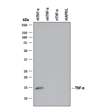


 下载产品说明书 下载SDS
 用小程序,查商品更便捷


 收藏
收藏
 对比
对比 咨询
咨询Immunocytochemistry(8-25 µg/mL)
ELISA Capture (Matched Antibody Pair)(2-8 µg/mL )
ELISA Detection (Matched Antibody Pair)(0.1-0.4 µg/mL )

Gly57-Leu233 (predicted)
Accession # P01375
Scientific Data
 View Larger
Detection of Recombinant Human TNF‑ alpha by Western Blot. Western blot shows 25 ng of Recombinant Human TNF-a (Catalog # 210-TA), Recombinant Mouse TNF-a aa 80-235 (Catalog # 410-MT), Recombinant Rat TNF-a (Catalog # 510-RT), and Recombinant Human APRIL/TNFSF13 (Catalog # 5860-AP). PVDF Membrane was probed with 1 µg/mL of Mouse Anti-Human TNF-a Monoclonal Antibody (Catalog # MAB610) followed by HRP-conjugated Anti-Mouse IgG Secondary Antibody (Catalog # HAF007). A specific band was detected for TNF-a at approximately 15 kDa (as indicated). This experiment was conducted under reducing conditions and using Immunoblot Buffer Group 3.
.") View Larger
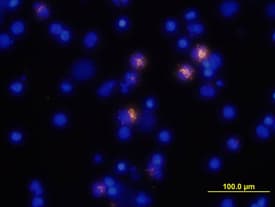
View LargerTNF‑ alpha in Human PBMCs. TNF-a was detected in immersion fixed human peripheral blood mononuclear cells (PBMCs) stimulated with LPS and monensin using Mouse Anti-Human TNF-a Monoclonal Antibody (Catalog # MAB610) at 10 µg/mL for 3 hours at room temperature. Cells were stained using the NorthernLights™ 557-conjugated Anti-Mouse IgG Secondary Antibody (yellow; Catalog # NL007) and counterstained with DAPI (blue). View our protocol for Fluorescent ICC Staining of Non-adherent Cells.
 View Larger
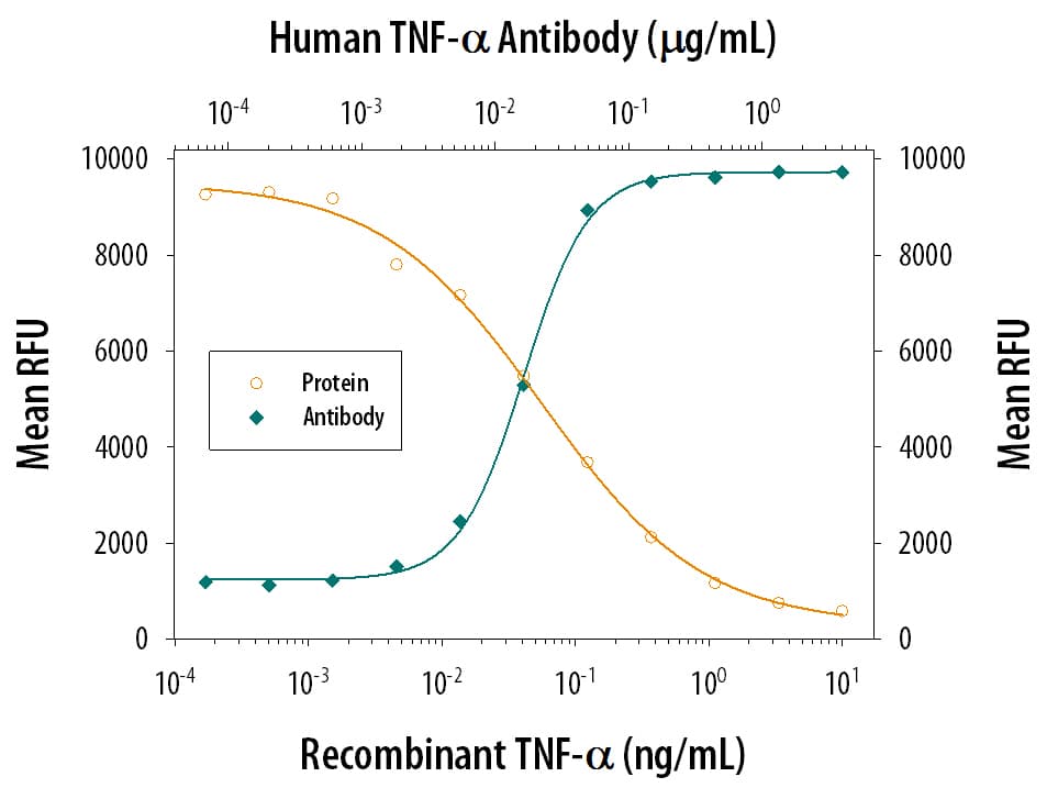
Cytotoxicity Induced by TNF‑ alpha and Neutralization by Human TNF‑ alpha Antibody. Recombinant Human TNF-a (Catalog # 210-TA) induces cytotoxicity in the the L-929 mouse fibroblast cell line in a dose-dependent manner (orange line). Cytotoxicity elicited by Recombinant Human TNF-a (0.25 ng/mL) is neutral-ized (green line) by increasing concentrations of Mouse Anti-Human TNF-a Monoclonal Antibody (Catalog # MAB610). The ND50 is typically 0.01-0.04 µg/mL in the presence of the metabolic inhibitor actinomycin D.
Human TNF-alpha Antibody Summary
Gly57-Leu233 (predicted)
Accession # P01375
Applications
Human TNF-alpha Sandwich Immunoassay
Please Note: Optimal dilutions should be determined by each laboratory for each application. General Protocols are available in the Technical Information section on our website.
Immunocytochemistry(8-25 µg/mL)
ELISA Capture (Matched Antibody Pair)(2-8 µg/mL )
ELISA Detection (Matched Antibody Pair)(0.1-0.4 µg/mL )
Background: TNF-alpha
Tumor necrosis factor alpha (TNF-alpha, TNF- alpha, TNFA ), also known as Cachectin and TNFSF2, is the prototypic ligand of the TNF superfamily. It is a pleiotropic molecule that plays a central role in inflammation, immune system development, apoptosis, and lipid metabolism. TNF- is produced by several lymphoid cells as well as by astrocytes, endothelial cells, and smooth muscle cells. Human TNF-alpha consisits of a 35 amino acid (aa) cytoplasmic domain, a 21 aa transmembrane segment, and a 177 aa extracellular domain (ECD). Within the ECD, human TNF-alpha shares 97% aa sequence identity with rhesus and 71%-92% with bovine, canine, cotton rat, equine, feline, mouse, porcine, and rat TNF-alpha. TNF-alpha is produced by a wide variety of immune, epithelial, endothelial, and tumor cells. TNF-alpha is assembled intracellularly to form a noncovalently linked homotrimer which is expressed on the cell surface. Cell surface TNF-alpha can induce the lysis of neighboring tumor cells and virus infected cells, and it can generate its own downstream cell signaling following ligation by soluble TNFR I. Shedding of membrane bound TNF-alpha by TACE/ADAM17 releases the bioactive cytokine, a 55 kDa molecular weight soluble trimer of the TNF-alpha extracellular domain. TNF-alpha binds the ubiquitous 55-60 kDa TNF RI and the hematopoietic cell-restricted 80 kDa TNF RII, both of which are also expressed as homotrimers present on virtually all cell types. Both type I and type II receptors bind TNF-alpha with comparable affinity, although only TNF RI contains a cytoplasmic death domain which triggers the activation of apoptosis. Soluble forms of both types of receptors are released and can neutralize the biological activity of TNF-alpha.
Preparation and Storage
- 12 months from date of receipt, -20 to -70 °C as supplied.
- 1 month, 2 to 8 °C under sterile conditions after reconstitution.
- 6 months, -20 to -70 °C under sterile conditions after reconstitution.
参考图片
Cytotoxicity Induced by TNF‑ alpha and Neutralization by Human TNF‑ alpha Antibody. Recombinant Human TNF‑ alpha (Catalog #
210‑TA) induces cytotoxicity in the the L‑929 mouse fibroblast cell line in a dose-dependent manner (orange line). Cytotoxicity elicited by Recombinant Human TNF‑ alpha (0.75 ng/mL) is neutralized (green line) by increasing concentrations of Mouse Anti-Human TNF‑ alpha Monoclonal Antibody (Catalog # MAB610). The ND50 is typically
0.01‑0.04 µg/mL in the presence of the metabolic inhibitor actinomycin D (1 µg/mL).
TNF‑ alpha in Human PBMCs. TNF‑ alpha was detected in immersion fixed human peripheral blood mononuclear cells (PBMCs) stimulated with LPS and monensin using Mouse Anti-Human TNF‑ alpha Monoclonal Antibody (Catalog # MAB610) at 10 µg/mL for 3 hours at room temperature. Cells were stained using the NorthernLights™ 557-conjugated Anti-Mouse IgG Secondary Antibody (yellow; Catalog # NL007) and counterstained with DAPI (blue). View our protocol for Fluorescent ICC Staining of Non-adherent Cells.