


 全部商品分类
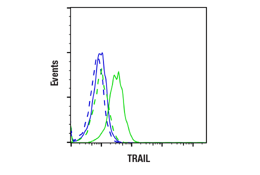


 用小程序,查商品更便捷


Monoclonal antibody is produced by immunizing animals with a synthetic peptide corresponding to residues surrounding Lys60 of human TRAIL, within the extracellular region of the protein.
Product Usage Information
| Application | Dilution |
|---|---|
| Western Blotting | 1:1000 |
| Simple Western™ | 1:10 - 1:50 |
| Immunoprecipitation | 1:50 |
| Immunohistochemistry (Paraffin) | 1:800 |
| Immunofluorescence (Immunocytochemistry) | 1:400 |
| Flow Cytometry (Fixed/Permeabilized) | 1:50 |
| Flow Cytometry (Live) | 1:50 |
Specificity/Sensitivity
Species Reactivity:
Human
Supplied in 10 mM sodium HEPES (pH 7.5), 150 mM NaCl, 100 µg/ml BSA, 50% glycerol and less than 0.02% sodium azide. Store at –20°C. Do not aliquot the antibody.
For a carrier free (BSA and azide free) version of this product see product #48318.
参考图片
Flow cytometric analysis of live THP-1 cells treated with TPA (80 nM, 18 h), rested for 48 h in fresh media, and then untreated (blue, negative) or treated with LPS (5 μg/mL, 18 h) (green, positive) and preincubated with human Fc block (2.5 μg/million cells, 10 min) using TRAIL (C92B9) Rabbit mAb (solid lines) or concentration-matched Rabbit (DA1E) mAb IgG XP® Isotype Control #3900 (dashed lines). Anti-rabbit IgG (H+L), F(ab')₂ Fragment (Alexa Fluor® 488 Conjugate) #4412 was used as a secondary antibody.
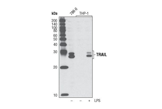
Western blot analysis of extract from 786-0 cells and differentiated THP-1 cells, untreated or treated overnight with LPS, using TRAIL (C92B9) Rabbit mAb.
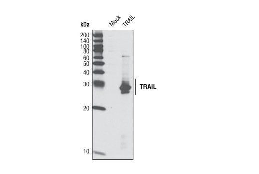
Western blot analysis of extracts from HeLa cells, mock transfected or transfected with human TRAIL expression construct, using TRAIL (C92B9) Rabbit mAb.
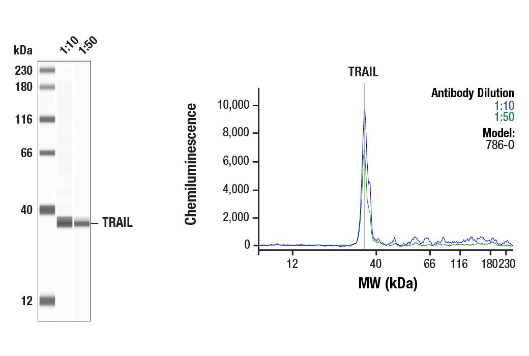
Simple Western™ analysis of 786-O (0.1 mg/mL) cell lysate using TRAIL (C92B9) Rabbit mAb #3219. The virtual lane view (left) shows the target band (as indicated) at 1:10 and 1:50 dilutions of primary antibody. The corresponding electropherogram view (right) plots chemiluminescence by molecular weight along the capillary at 1:10 (blue line) and 1:50 (green line) dilutions of primary antibody. This experiment was performed under reducing conditions on the Jess™ Simple Western instrument from ProteinSimple, a BioTechne brand, using the 12-230 kDa separation module.
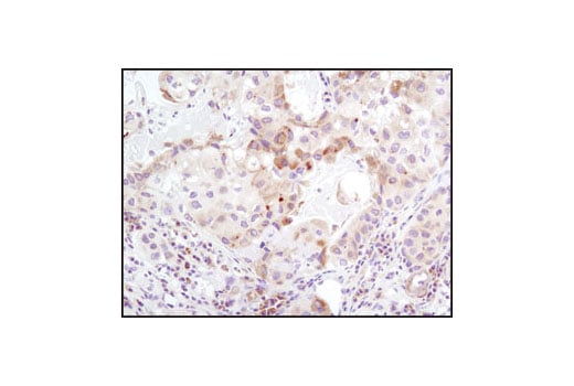
Immunohistochemical analysis of paraffin-embedded human lung carcinoma using TRAIL (C92B9) Rabbit mAb.
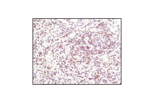
Immunohistochemical analysis of paraffin-embedded human lymphoma using TRAIL (C92B9) Rabbit mAb.
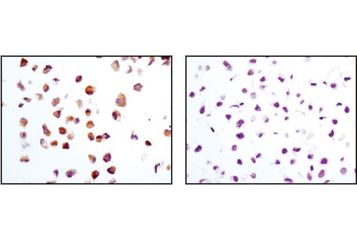
Immunohistochemical analysis of paraffin-embedded 786-0 (positive, left) or HeLa (negative, right) cell pellets using TRAIL (C92B9) Rabbit mAb.
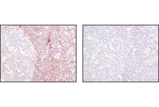
Immunohistochemical analysis of paraffin-embedded human tonsil using TRAIL (C92B9) Rabbit mAb in the presence of control peptide (left) or antigen-specific peptide (right).
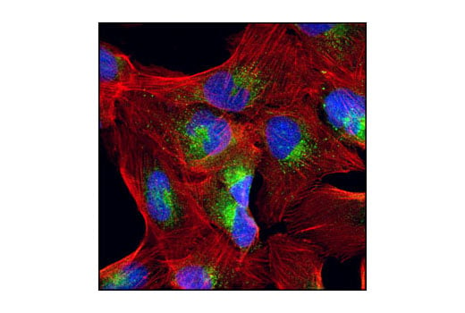
Confocal immunofluorescent analysis of 786-0 cells using TRAIL (C92B9) Rabbit mAb (green). Actin filaments have been labeled with DY-554 phalloidin (red). Blue pseudocolor = DRAQ5™ (fluorescent DNA dye).
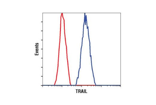
Flow cytometric analysis of 786-0 cells using TRAIL (C92B9) Rabbit mAb (blue) compared to a nonspecific negative control antibody (red).