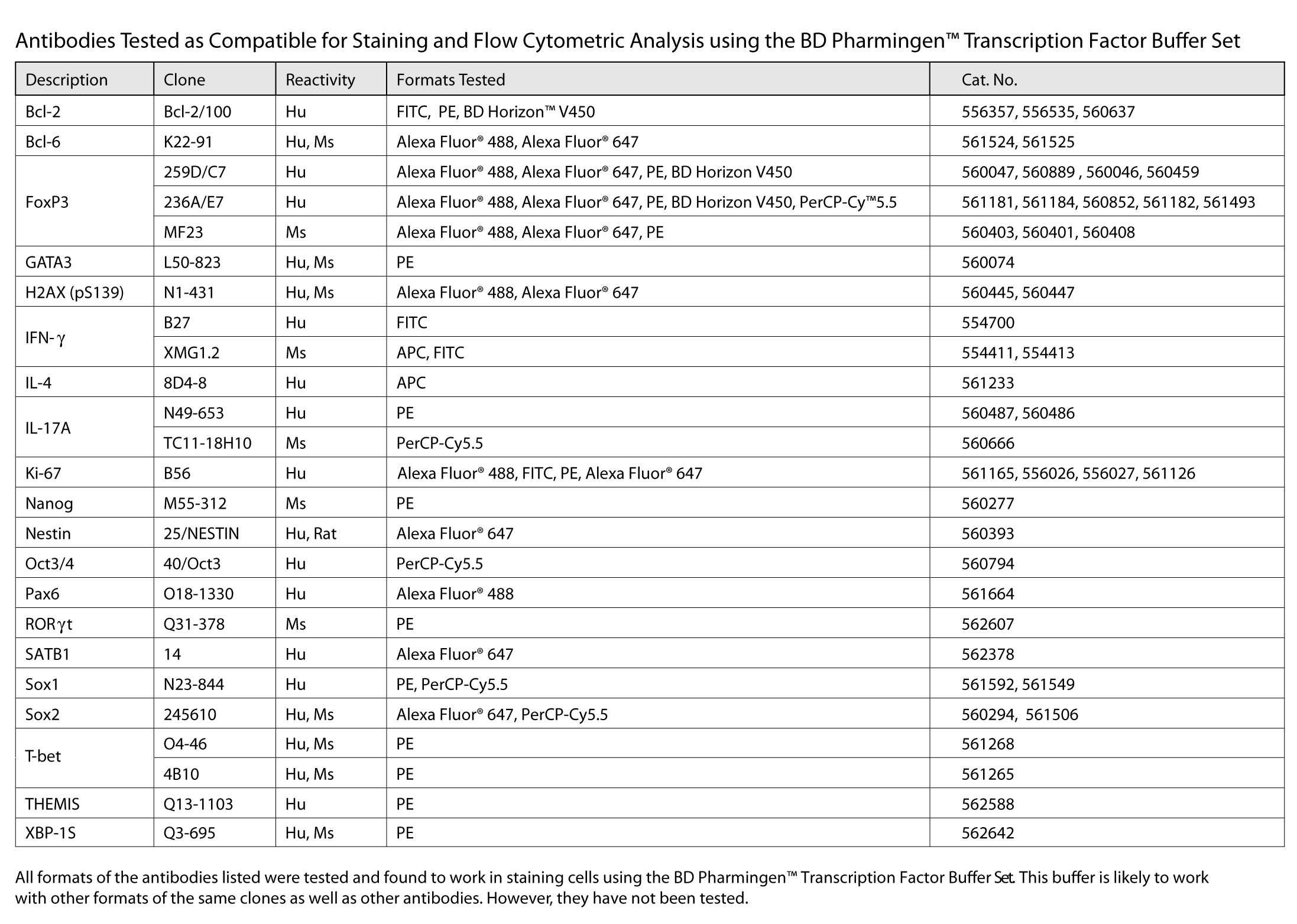
The BD Pharmingen™ Transcription Factor Buffer Set is optimized for fixing and permeabilizing cells prior to immunofluorescent staining and flow cytometric analysis of cells that express specific intracytoplasmic and intranuclear proteins. The BD Pharmingen™ Transcription Factor Buffer Set was designed to improve ease-of-use and minimize processing time, to reduce nonspecific staining, to increase the resolution of positively stained cells and to significantly reduce cell loss during fixation, permeabilization and staining procedures. Flow cytometric detection of the many proteins known to be expressed within various intracellular compartments, especially transcription factors, is improved with BD Pharmingen™ Transcription Factor Buffer Set use. This buffer system has been found useful for fixing and permeabilizing a variety of cell types from diverse human and mouse tissues. The buffer system is flexible in supporting multiwell-plate high-throughput and bulk sample analyses and applications that require overnight sample storage. The buffer system has minimal impact on the light-scatter and autofluorescence characteristics of processed cells resulting in characteristics similar to those observed for freshly prepared, highly viable primary cells. In many cases the buffer system was found to be compatible with the immunofluorescent staining of cell-surface antigens both before and after cellular fixation and permeabilization. The buffer system is also compatible with many tandem fluorochromes.
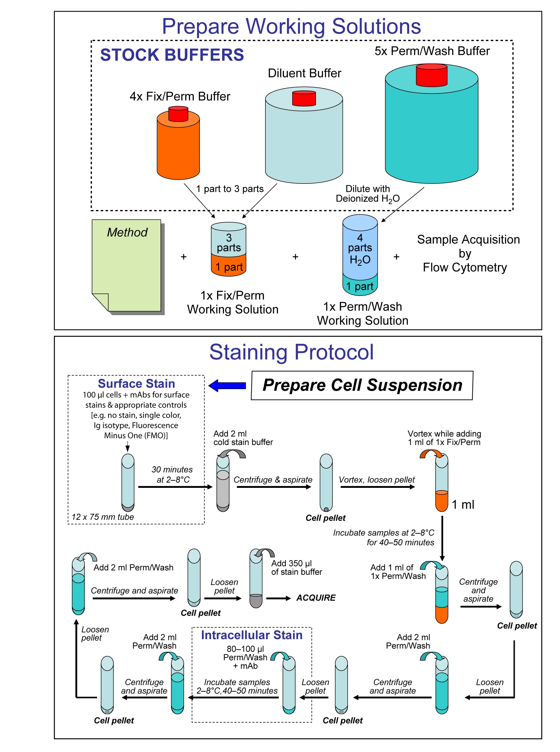
Overview of Buffer Dilution and Staining Protocol.
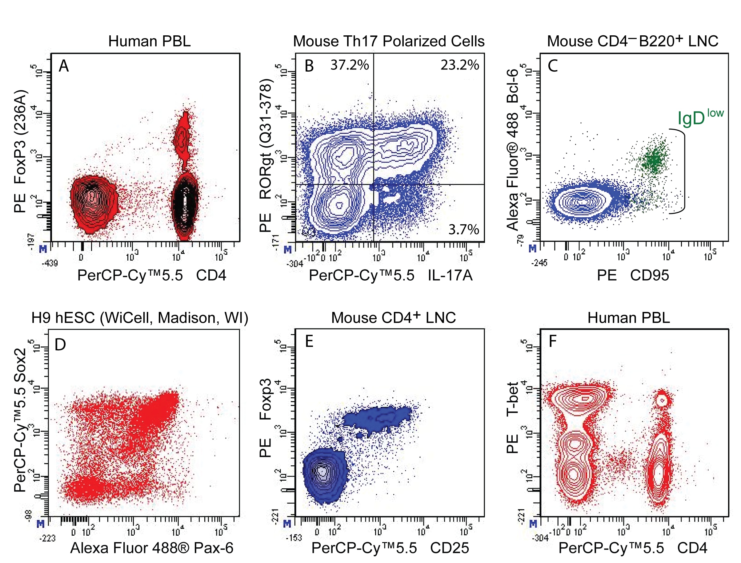
Multicolor flow cytometric analysis of transcription factors expressed in different cell types using the BD Pharmingen™ Transcription Factor Buffer Set. Bivariate flow cytometric plots showing (A) CD4 versus FoxP3 expression in human peripheral blood lymphocytes (PBL); (B) IL-17A versus RORgt expression in BALB/c mouse Th17-polarized cells; (C) CD95 versus Bcl-6 expression in C57BL/6 mouse lymph node cells (LNC) and identification of germinal center B-cells using CD4-B220+IgDloCD95hi phenotype as green colorized dots; (D) Pax-6 versus Sox-2 in H9 (WiCell, Madison, Wi) human embryonic stem cell (ESC) derived neural cultures; (E) mouse CD25 versus Foxp3 expression in CD4 T cells derived from C57BL/6 mouse LNC; (F) CD4 versus T-bet expression in human PBL. Plots were derived from gated events with the forward and side light-scattering characteristics of intact lymphocytes or indicated cell types using BD FACSDiva™ Software v. 6.1.3 and a BD LSRFortessa™ Flow Cytometer System.


 全部商品分类


 下载产品说明书
 用小程序,查商品更便捷


 收藏
收藏
 对比
对比 咨询
咨询