


 全部商品分类
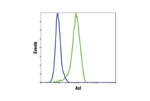


 用小程序,查商品更便捷


Monoclonal antibody is produced by immunizing animals with a recombinant human fragment of the Axl protein.
Product Usage Information
| Application | Dilution |
|---|---|
| Western Blotting | 1:1000 |
| Simple Western™ | 1:10 - 1:50 |
| Immunoprecipitation | 1:50 |
| Immunohistochemistry (Paraffin) | 1:300 - 1:1200 |
| Immunofluorescence (Immunocytochemistry) | 1:100 - 1:400 |
| Flow Cytometry (Fixed/Permeabilized) | 1:100 - 1:400 |
Specificity/Sensitivity
Species Reactivity:
Human, Monkey
Supplied in 10 mM sodium HEPES (pH 7.5), 150 mM NaCl, 100 µg/ml BSA, 50% glycerol and less than 0.02% sodium azide. Store at –20°C. Do not aliquot the antibody.
For a carrier free (BSA and azide free) version of this product see product #80287.
参考图片
Flow cytometric analysis of fixed and permeabilized Jurkat cells (blue, negative) and DU145 cells (green, positive) using Axl (C89E7H4) Rabbit mAb. Anti-rabbit IgG (H+L), F(ab')2 Fragment (Alexa Fluor® 488 Conjugate) #4412 was used as a secondary antibody.
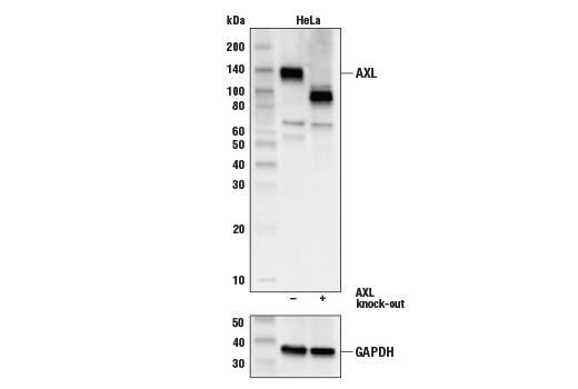
Western blot analysis of HeLa Cell Extracts, untreated (-) or Axl knock-out (+) using Axl (C89E7) Rabbit mAb, #8661 (upper) or GAPDH (D16H11) XP® Rabbit mAb, #5174 (lower).
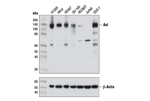
Western blot analysis of extracts from various cell lines using Axl (C89E7) Rabbit mAb (upper) and β-Actin (D6A8) Rabbit mAb #8457 (lower).
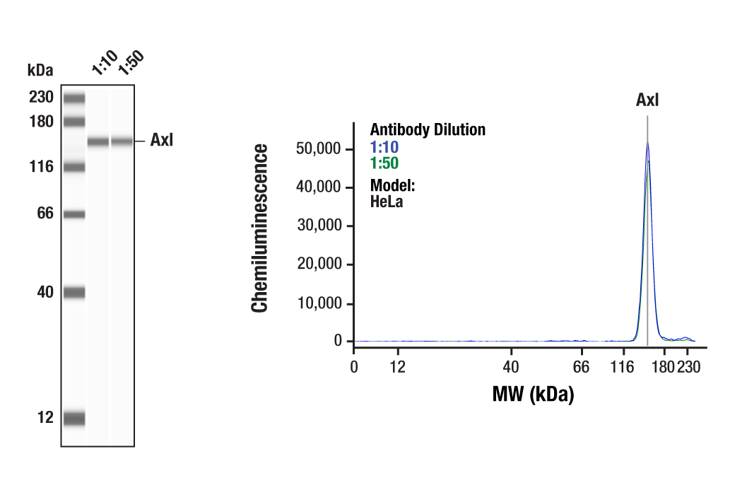
Simple Western™ analysis of lysates (0.1 mg/mL) from HeLa cells using Axl (C89E7) Rabbit mAb #8661. The virtual lane view (left) shows a single target band (as indicated) at 1:10 and 1:50 dilutions of primary antibody. The corresponding electropherogram view (right) plots chemiluminescence by molecular weight along the capillary at 1:10 (blue line) and 1:50 (green line) dilutions of primary antibody. This experiment was performed under reducing conditions on the Jess™ Simple Western instrument from ProteinSimple, a BioTechne brand, using the 12-230 kDa separation module.
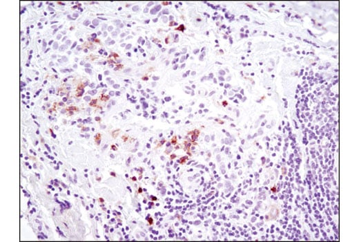
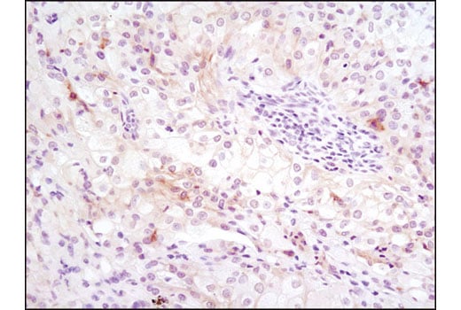
Immunohistochemical analysis of paraffin-embedded breast carcinoma using Axl (C89E7) Rabbit mAb. Note staining of infiltrating cells.
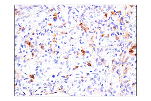
Immunohistochemical analysis of paraffin-embedded human renal cell carcinoma using Axl (C89E7) Rabbit mAb.
Immunohistochemical analysis of paraffin-embedded human B-cell non-Hodgkin's lymphoma using Axl (C89E7) Rabbit mAb.
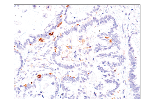
Immunohistochemical analysis of paraffin-embedded human ovarian serous carcinoma using Axl (C89E7) Rabbit mAb.
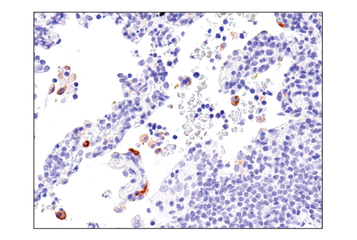
Immunohistochemical analysis of paraffin-embedded metastatic lung carcinoma using Axl (C89E7) Rabbit mAb.
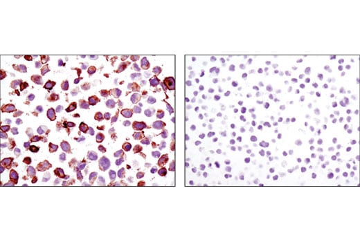
Immunohistochemical analysis of paraffin-embedded cell pellets, NCI-H1299 (left) or Jurkat (right), using Axl (C89E7) Rabbit mAb.
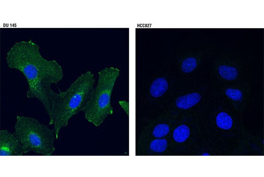
Confocal immunofluorescent analysis of DU 145 (left) and HCC827 (right) cells using Axl (C89E7) Rabbit mAb (green). Blue pseudocolor = DRAQ5® #4084 (fluorescent DNA dye).