

 全部商品分类
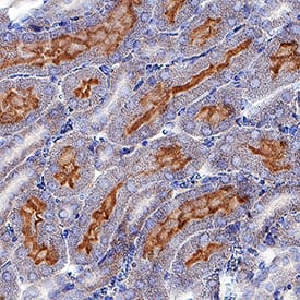


 用小程序,查商品更便捷


Scientific Data
.") View Larger
View LargerAxl in Mouse Kidney. Axl was detected in immersion fixed paraffin-embedded sections of mouse kidney using Goat Anti-Mouse Axl Antigen Affinity-purified Polyclonal Antibody (Catalog # AF854) at 3 µg/mL for 1 hour at room temperature followed by incubation with the Anti-Goat IgG VisUCyte™ HRP Polymer Antibody (Catalog # VC004). Before incubation with the primary antibody, tissue was subjected to heat-induced epitope retrieval using Antigen Retrieval Reagent-Basic (Catalog # CTS013). Tissue was stained using DAB (brown) and counterstained with hematoxylin (blue). Specific staining was localized to cell membranes in convoluted tubules. View our protocol for IHC Staining with VisUCyte HRP Polymer Detection Reagents.
Mouse Axl Antibody Summary
His20-Pro443
Accession # Q80YQ3
Applications
Please Note: Optimal dilutions should be determined by each laboratory for each application. General Protocols are available in the Technical Information section on our website.
Background: Axl
Axl (Ufo, Ark), Dtk (Sky, Tyro3, Rse, Brt), and Mer (human and mouse homologues of chicken c-Eyk) constitute a subfamily of the receptor tyrosine kinases (1, 2). The extracellular domains of these proteins contain two Ig-like motifs and two fibronectin type III motifs. This characteristic topology is also found in neural cell adhesion molecules and in receptor tyrosine phosphatases. The mouse Axl cDNA encodes an 888 amino acid (aa) precursor that includes an 18 aa signal sequence, a 427 aa extracellular domain, a 21 aa transmembrane segment, and a 422 aa cytoplasmic domain. The extracellular domains of mouse and human Axl share 81% aa sequence identity. These receptors bind the vitamin K-dependent protein growth arrest specific gene 6 (Gas6) which is structurally related to the anticoagulation factor protein S. Binding of Gas6 induces receptor autophosphorylation and downstream signaling pathways that can lead to cell proliferation, migration, or the prevention of apoptosis (3). This family of tyrosine kinase receptors is involved in hematopoiesis, embryonic development, tumorigenesis, and regulation of testicular functions.
- Yanagita, M. (2004) Curr. Opin. Nephrol. Hypertens. 13:465.
- Nagata, K. et al. (1996) J. Biol. Chem. 22:30022.
- Holland, S. et al. (2005) Canc. Res. 65:9294.
Preparation and Storage
- 12 months from date of receipt, -20 to -70 °C as supplied.
- 1 month, 2 to 8 °C under sterile conditions after reconstitution.
- 6 months, -20 to -70 °C under sterile conditions after reconstitution.