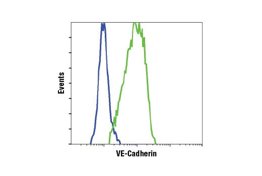
VE-Cadherin (D87F2) XP ®  Rabbit mAb

VE-Cadherin (D87F2) XP ® Rabbit mAb

分享
品牌: CST
pdf 下载产品说明书
pdf 下载SDS
用小程序,查商品更便捷
收藏
对比对比
咨询咨询
    分子量:
    130-140
    130-140
    反应种属:
    Human,Bovine,Pig,
    Human,Bovine,Pig,
    展开
    产品介绍
    产品信息
    抗原名称
    VE-Cadherin
    来源纯化
    使用与人血管内皮钙粘素的羧基末端周围的残基相对应的合成肽,对动物进行免疫接种来产生单克隆抗体。
    宿主
    Rabbit
    简单描述
    Monoclonal Antibody for studying VE-Cadherin. Cited in 247 publications. Validated for WB, IP, IF, F. Available in 2 sizes. Highly specific and rigorously validated in-house, VE-Cadherin (D87F2) XP® Rabbit Monoclonal Antibody (CST #2500) is ready to ship.
    商品描述

    Product Usage Information

    ApplicationDilution
    Western Blotting1:1000
    Immunoprecipitation1:50
    Immunofluorescence (Immunocytochemistry)1:400
    Flow Cytometry (Fixed/Permeabilized)1:100
    分子量
    130-140
    研究领域
    癌症,细胞生物学,纤维化,神经科学,
    应用
    反应种属
    Human,Bovine,Pig,
    目标/特异性

    Specificity/Sensitivity

    VE-Cadherin (D87F2) XP® Rabbit mAb 可识别内源水平的总血管内皮钙粘素蛋白。该抗体不与其他钙粘素家族蛋白发生交叉反应。

    物种反应性:

    人, 牛 , 猪

    敏感性
    Endogenous
    背景
    背景
    钙黏着蛋白是在其胞外结构域中含有大约 100 个残基的钙黏着蛋白重复序列的跨膜糖蛋白超家族。钙黏着蛋白介导钙依赖性细胞-细胞黏附,并在正常组织发育中发挥至关重要的作用 (1)。经典的钙黏着蛋白亚家族包括 N-、P-、R-、B- 和 E-钙黏着蛋白,以及约十个在黏附连接中存在的其他成员,黏附连接是一种在极化上皮细胞顶面的细胞结构。经典钙黏着蛋白的胞质结构域可与 β-联蛋白、γ-联蛋白(也称作斑珠蛋白)和 p120 联蛋白相互作用。β-联蛋白和 γ-联蛋白与 α-联蛋白结合, 并且 α-联蛋白将钙黏着蛋白-联蛋白复合体连接至肌动蛋白细胞骨架 (1,2)。β- 和 γ-联蛋白在连接复合体中发挥结构性作用,而 p120 可调节钙黏着蛋白黏附活性和转运 (1-4)。研究者认为 E-钙黏着蛋白是许多上皮癌侵润和生长的活性抑制蛋白 (1-3)。研究表明,除了上皮钙粘素丢失外,癌细胞的神经钙粘素表达上调。钙粘素表达的这种变化被称作“钙粘素转换”。N-钙黏着蛋白与 FGF 受体协作,导致 MMP-9 过表达和细胞侵袭 (3)。研究表明,在内皮细胞中,VE-钙黏着蛋白信号转导、表达和定位与血管通透性和肿瘤血管生成相关 (5,6)。研究者还已经证实,正常情况下在上皮细胞中的存在的 P-钙黏着蛋白,其表达在卵巢和其他人类癌症中也发生改变 (7,8)。 1.Wheelock, M.J. and Johnson, K.R. (2003) Annu Rev Cell Dev Biol 19, 207-35. 2.Christofori, G. (2003) EMBO J 22, 2318-23. 3.Hazan, R.B. et al. (2004) Ann N Y Acad Sci 1014, 155-63. 4.Bryant, D.M. and Stow, J.L. (2004) Trends Cell Biol 14, 427-34. 5.Rabascio, C. et al. (2004) Cancer Res 64, 4373-7. 6.Yamaoka-Tojo, M. et al. (2006) Arterioscler Thromb Vasc Biol 26, 1991-7. 7.Patel, I.S. et al. (2003) Int J Cancer 106, 172-7. 8.Sanders, D.S. et al. (2000) J Pathol 190, 526-30.
    研究领域
    癌症,细胞生物学,纤维化,神经科学,
    翻译后修饰
    Unmodified
    制备和贮存
    保存方式
    Supplied in 10 mM sodium HEPES (pH 7.5), 150 mM NaCl, 100 µg/ml BSA, 50% glycerol and less than 0.02% sodium azide. Store at –20°C. Do not aliquot the antibody.
    数据库链接
    Entrez-Gene ID
    1003
    UniProt ID
    P33151

    参考图片

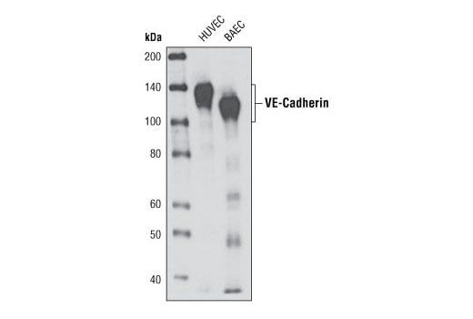
    Western blot analysis of extracts from HUVEC and BAEC cells using VE-Cadherin (D87F2) XP® Rabbit mAb.使用VE-Cadherin (D87F2) 兔单抗对HUVEC和BAEC细胞提取物进行western blot分析。

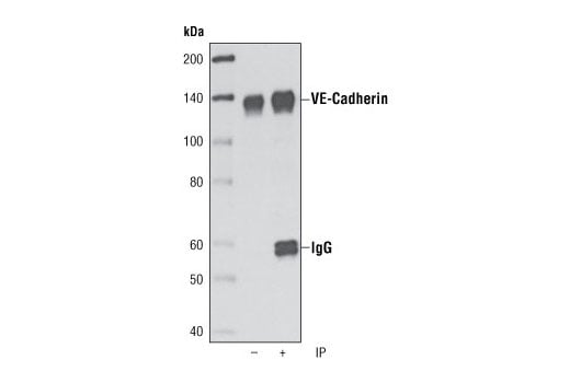
    Immunoprecipitation of VE-caderin from HUVEC cells using VE-Cadherin (D87F2) XP® Rabbit mAb followed by western blot using the same antibody. Lane 1 is 5% input.使用VE-Cadherin (D87F2) 兔单抗对HUVEC细胞的VE-caderin进行免疫共沉淀实验,随后使用同样的抗体进行western blot分析。第一道为5%上样。

    Flow cytometric analysis of HeLa cells (blue) and HUVEC cells (green) using VE-Cadherin (D87F2) Rabbit mAb.使用VE-Cadherin (D87F2) 兔单抗对HeLa细胞(蓝色)和HUVEC细胞(绿色)进行流式细胞分析。

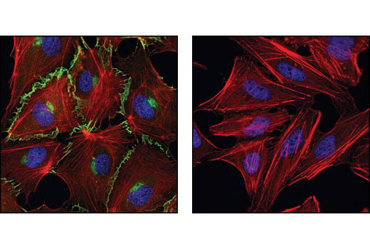
    Confocal immunofluorescent analysis of HUVE cells (left) and HeLa cells (right) using VE-Cadherin (D87F2) XP® Rabbit mAb (green). Actin filaments have been labeled with DY-554 phalloidin (red). Blue pseudocolor = DRAQ5® #4084 (fluorescent DNA dye).使用VE-Cadherin (D87F2) 兔单抗对HUVE细胞(左)和HeLa细胞(右)进行激光共聚焦免疫荧光分析。肌动蛋白丝使用DY-554鬼笔环肽标记(红色)。蓝色假色=DRAQ5® #4084 (DNA荧光染料)。

    声明 :本官网所有报价均为常温或者蓝冰运输价格,如有产品需要干冰运输,需另外加收干冰运输费。
    货号:
    2500T
    一键复制
    询价
    供应商现货
    20ul
    100ul
    抗体送U盘
    详情
    选择数量
    当前规格1件起购 
    配送至
    预计5-6个工作日送达,快递: 免运费,若需干冰额外收费