


 全部商品分类
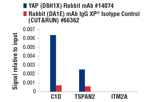


 用小程序,查商品更便捷


Monoclonal antibody is produced by immunizing animals with recombinant protein specific to the carboxy terminus of human YAP protein. The epitope corresponds to a region surrounding Pro435 of human YAP isoform 1. This sequence region is 100% conserved among all known isoforms of human YAP protein.
Product Usage Information
For optimal ChIP and ChIP-seq results, use 10 μl of antibody and 10 μg of chromatin (approximately 4 x 10⁶ cells) per IP. This antibody has been validated using SimpleChIP® Enzymatic Chromatin IP Kits.
The CUT&RUN dilution was determined using CUT&RUN Assay Kit #86652.
| Application | Dilution |
|---|---|
| Western Blotting | 1:1000 |
| Simple Western™ | 1:10 - 1:50 |
| Immunoprecipitation | 1:50 |
| IHC Leica Bond | 1:100 - 1:400 |
| Immunohistochemistry (Paraffin) | 1:200 - 1:800 |
| Immunofluorescence (Frozen) | 1:50 - 1:200 |
| Immunofluorescence (Immunocytochemistry) | 1:50 - 1:200 |
| Flow Cytometry (Fixed/Permeabilized) | 1:50 - 1:200 |
| Chromatin IP | 1:50 |
| Chromatin IP-seq | 1:50 |
| CUT&RUN | 1:50 |
Specificity/Sensitivity
Species Reactivity:
Human, Mouse, Rat, Hamster, Monkey
Supplied in 10 mM sodium HEPES (pH 7.5), 150 mM NaCl, 100 µg/ml BSA, 50% glycerol and less than 0.02% sodium azide. Store at –20°C. Do not aliquot the antibody.
For a carrier free (BSA and azide free) version of this product see product #53921.
参考图片
CUT&RUN was performed with NCI-H2052 cells and either YAP (D8H1X) XP® Rabbit mAb or Rabbit (DA1E) mAb IgG XP® Isotype Control (CUT&RUN) #66362, using CUT&RUN Assay Kit #86652. The enriched DNA was quantified by real-time PCR using human C1D downstream primers, human TSPAN2 upstream primers, and human ITM2A upstream primers. The amount of immunoprecipitated DNA in each sample is represented as signal relative to the total amount of input chromatin, which is equivalent to one.
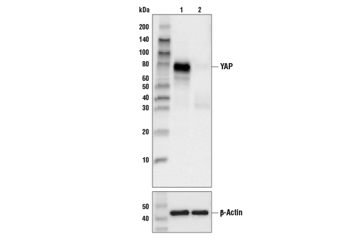
Western blot analysis of extracts from control HeLa cells (Lane 1) or YAP knockout HeLa cells (Lane 2) using YAP (D8H1X) XP® Rabbit mAb (upper) or β-actin (13E5) Rabbit mAb #4970 (lower). The absence of signal in the YAP knockout HeLa cells confirms the specificity of the antibody for YAP.
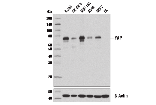
Western blot analysis of extracts from various cell lines using YAP (D8H1X) XP® Rabbit mAb (upper) and β-Actin (D6A8) Rabbit mAb #8457 (lower). As expected, RL cells are negative for YAP protein expression.
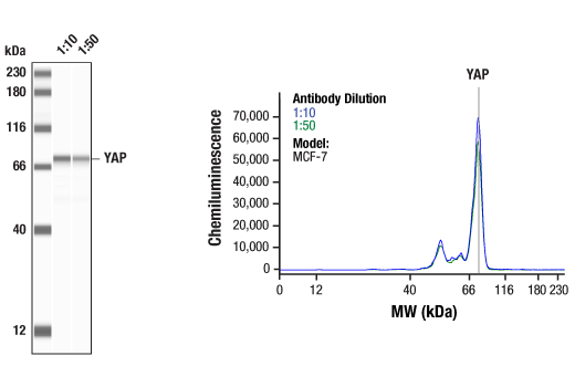
Simple Western™ analysis of lysates (1 mg/mL) from MCF-7 cells using YAP (D8H1X) XP® Rabbit mAb #14074. The virtual lane view (left) shows the target band (as indicated) at 1:10 and 1:50 dilutions of primary antibody. The corresponding electropherogram view (right) plots chemiluminescence by molecular weight along the capillary at 1:10 (blue line) and 1:50 (green line) dilutions of primary antibody. This experiment was performed under reducing conditions on the Jess™ Simple Western instrument from ProteinSimple, a BioTechne brand, using the 12-230 kDa separation module.
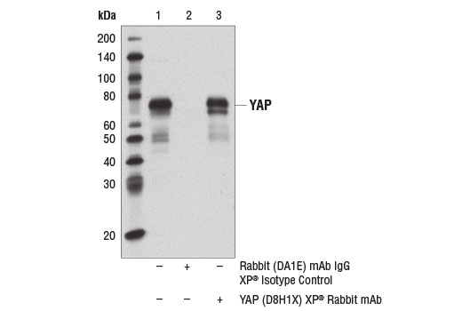
Immunoprecipitation of YAP protein from A-204 cell extracts using Rabbit (DA1E) mAb XP® Isotype Control #3900 (lane 2) or YAP (D8H1X) XP® Rabbit mAb (lane 3). Lane 1 is 10% input. Western blot analysis was performed using YAP (D8H1X) XP® Rabbit mAb. Mouse Anti-rabbit IgG (Conformation Specific) (L27A9) mAb #3678 was used as a secondary antibody.
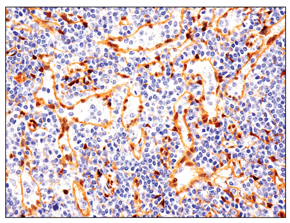
Immunohistochemical analysis of paraffin-embedded human non-Hodgkin lymphoma using YAP (D8H1X) XP® Rabbit mAb performed on the Leica® BOND™ Rx.
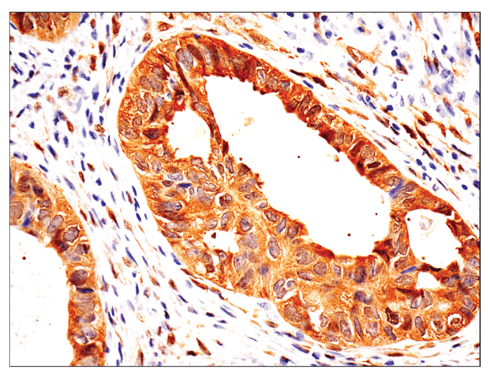
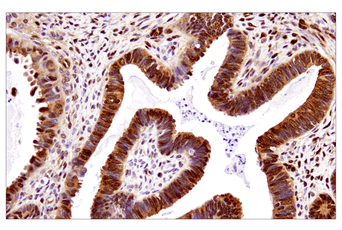
Immunohistochemical analysis of paraffin-embedded human endometrioid adenocarcinoma using YAP (D8H1X) XP® Rabbit mAb performed on the Leica® BOND™ Rx.
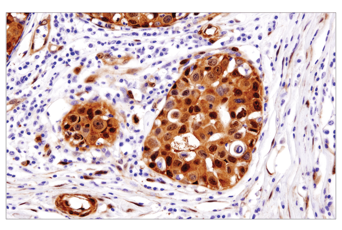
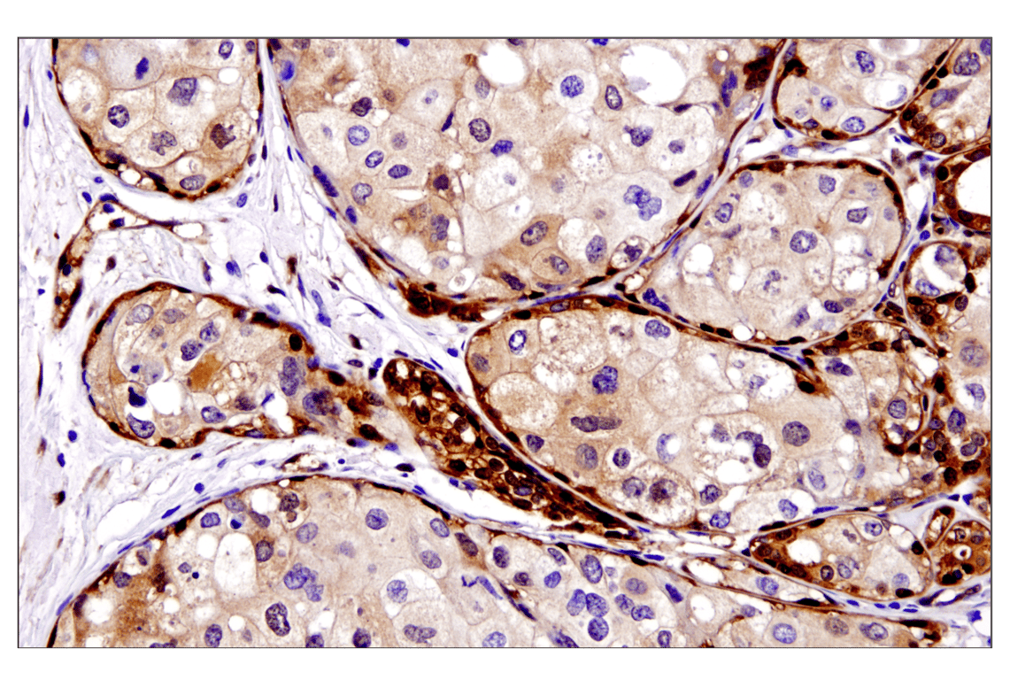
Immunohistochemical analysis of paraffin-embedded human breast adenocarcinoma using YAP (D8H1X) XP® Rabbit mAb.
Immunohistochemical analysis of paraffin-embedded human breast adenocarcinoma using YAP (D8H1X) XP® Rabbit mAb.
Immunohistochemical analysis of paraffin-embedded human ovarian serous carcinoma using YAP (D8H1X) XP® Rabbit mAb.
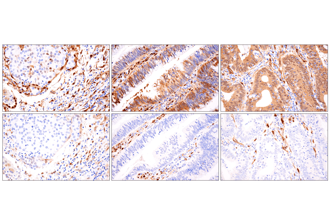
Immunohistochemical analysis of paraffin-embedded human non-small cell lung carcinoma (left), colon carcinoma case 1 (middle) or colon carcinoma case 2 (right), using YAP (D8H1X) XP® Rabbit mAb (top) or TAZ (E9J5A) XP® Rabbit mAb #72804 (bottom).
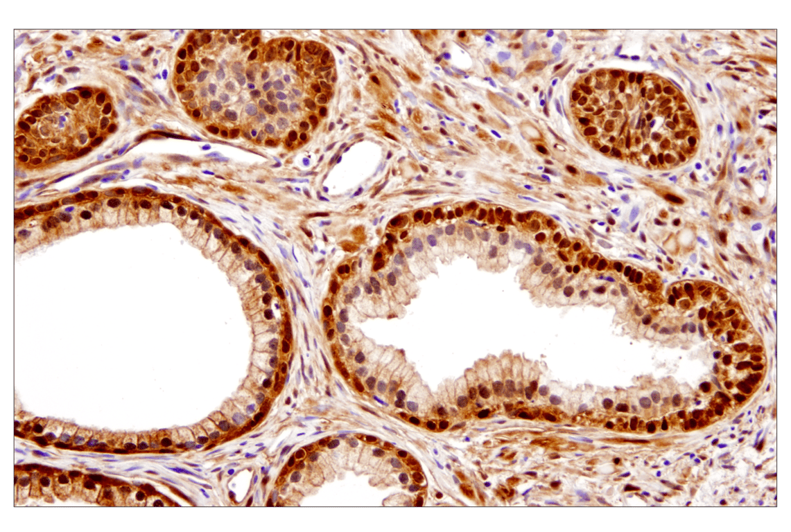
Immunohistochemical analysis of paraffin-embedded human benign prostatic hyperplasia using YAP (D8H1X) XP® Rabbit mAb.
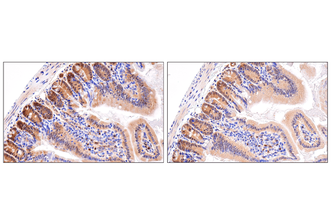
Immunohistochemical analysis of paraffin-embedded mouse small intesine using YAP (D8H1X) XP® Rabbit mAb (left) or YAP Antibody (right). These two antibodies detect unique, non-overlapping epitopes on YAP protein. The similar staining patterns obtained with both antibodies help to confirm the specificity of the staining.
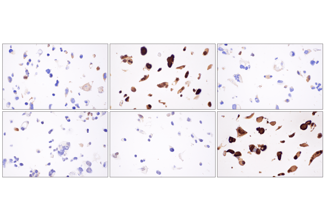
Immunohistochemical analysis of paraffin-embedded BEN cell pellet, untransfected (left, YAP/TAZ low), YAP-transfected (middle) or TAZ-transfected (right), using YAP (D8H1X) XP® Rabbit mAb (top) or TAZ (E9J5A) XP® Rabbit mAb #72804 (bottom).
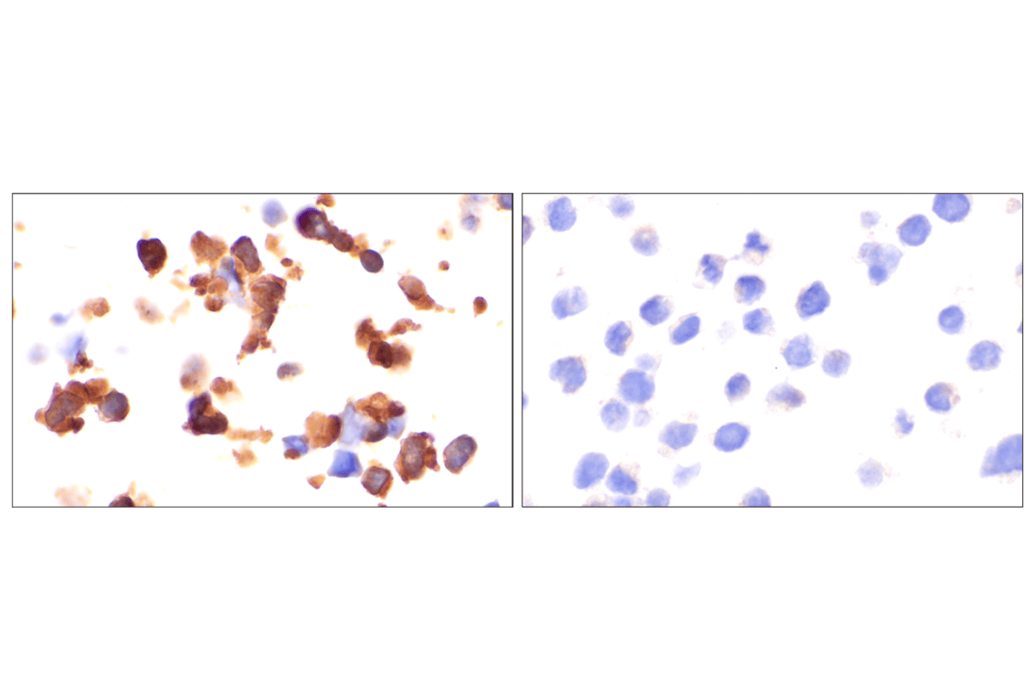
Immunohistochemical analysis of paraffin-embedded 293T cell pellet, wild-type (left, positive) or YAP/TAZ knockout (right, negative), using YAP (D8H1X) XP® Rabbit mAb.
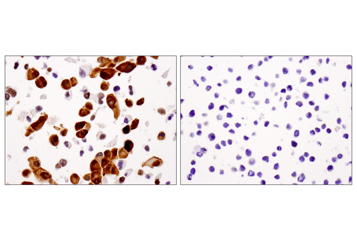
Immunohistochemical analysis of paraffin-embedded PANC-1 (left) and RL (right) cell pellets using YAP (D8H1X) XP® Rabbit mAb.
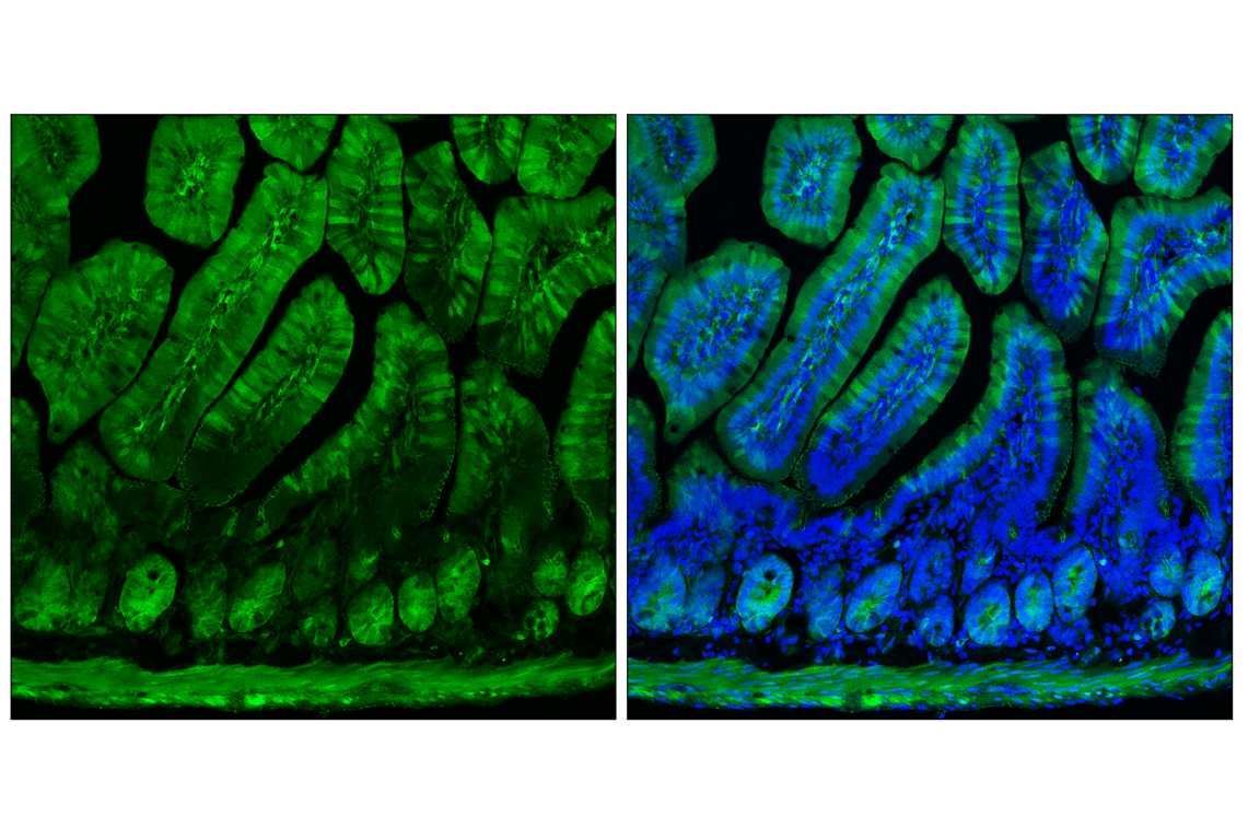
Confocal immunofluorescent analysis of fixed frozen mouse small intestine labeled with YAP (D8H1X) XP® Rabbit mAb (left, green) and co-labeled with ProLong® Gold Antifade Reagent with DAPI #8961 (right, blue).
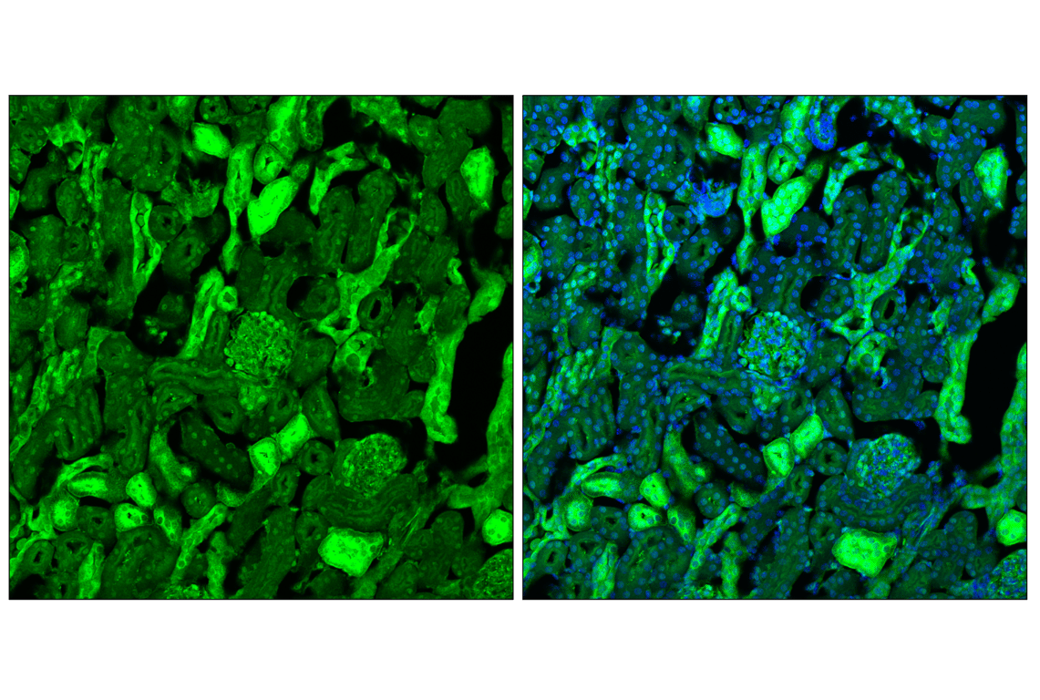
Confocal immunofluorescent analysis of fixed frozen mouse kidney labeled with YAP (D8H1X) XP® Rabbit mAb (left, green) and co-labeled with ProLong® Gold Antifade Reagent with DAPI #8961 (right, blue).
Confocal immunofluorescent analysis of fixed frozen mouse cerebellum labeled with YAP (D8H1X) XP® Rabbit mAb (left, green) and co-labeled with ProLong® Gold Antifade Reagent with DAPI #8961 (right, blue).
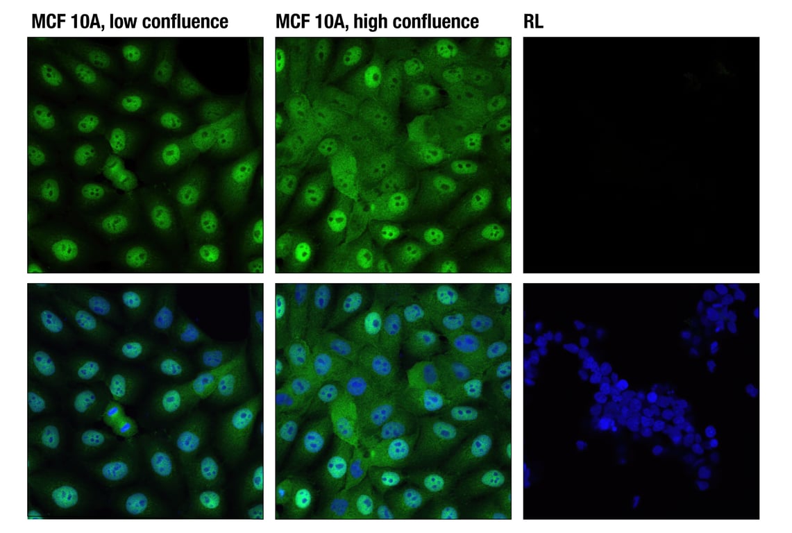
Confocal immunofluorescent analysis of low confluence MCF 10A cells (left), high confluence MCF 10A (center), and YAP negative RL cells (right) using YAP (D8H1X) XP® Rabbit mAb (green). Blue pseudocolor in lower images = DRAQ5® #4084 (fluorescent DNA dye). Increased nuclear localization of YAP protein is seen in low confluence (proliferating) cells.
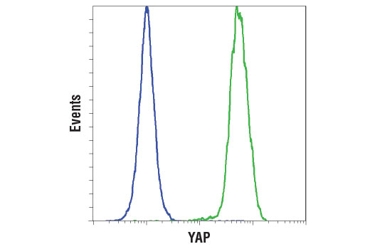
Flow cytometric analysis of RL cells (blue) and A-204 cells (green) using YAP (D8H1X) XP® Rabbit mAb. Anti-rabbit IgG (H+L), F(ab')2 Fragment (Alexa Fluor® 488 Conjugate) #4412 was used as a secondary antibody.
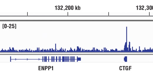
Chromatin immunoprecipitations were performed with cross-linked chromatin from NCI-H2052 cells and YAP (D8H1X) XP® Rabbit mAb, using SimpleChIP® Enzymatic Chromatin IP Kit (Magnetic Beads) #9003. DNA Libraries were prepared using DNA Library Prep Kit for Illumina® (ChIP-seq, CUT&RUN) #56795. The figure shows binding across CTGF, a known target gene of YAP (see additional figure containing ChIP-qPCR data).
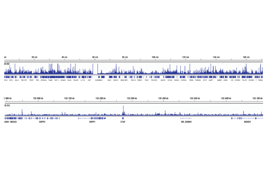
Chromatin immunoprecipitations were performed with cross-linked chromatin from NCI-H2052 cells and YAP (D8H1X) XP® Rabbit mAb, using SimpleChIP® Enzymatic Chromatin IP Kit (Magnetic Beads) #9003. DNA Libraries were prepared using DNA Library Prep Kit for Illumina® (ChIP-seq, CUT&RUN) #56795. The figure shows binding across chromosome 6 (upper), including CTGF (lower), a known target gene of YAP (see additional figure containing ChIP-qPCR data).
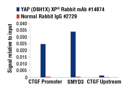
Chromatin immunoprecipitations were performed with cross-linked chromatin from NCI-2052 cells and either YAP (D8H1X) XP® Rabbit mAb, or Normal Rabbit IgG #2729, using SimpleChIP® Enzymatic Chromatin IP Kit (Magnetic Beads) #9003. The enriched DNA was quantified by real-time PCR using SimpleChIP® Human CTGF Promoter Primers #14927, human SMYD3 intron 2 primers, and SimpleChIP® Human CTGF Upstream Primers #14928. The amount of immunoprecipitated DNA in each sample is represented as signal relative to the total amount of input chromatin, which is equivalent to one.
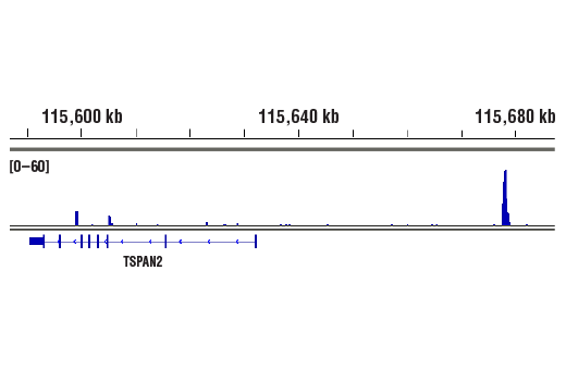
CUT&RUN was performed with NCI-H2052 cells and YAP (D8H1X) XP® Rabbit mAb, using CUT&RUN Assay Kit #86652. DNA libraries were prepared using DNA Library Prep Kit for Illumina® (ChIP-seq, CUT&RUN) #56795. The figure shows binding around TSPAN2, a known target gene of YAP (see additional figure containing CUT&RUN-qPCR data).
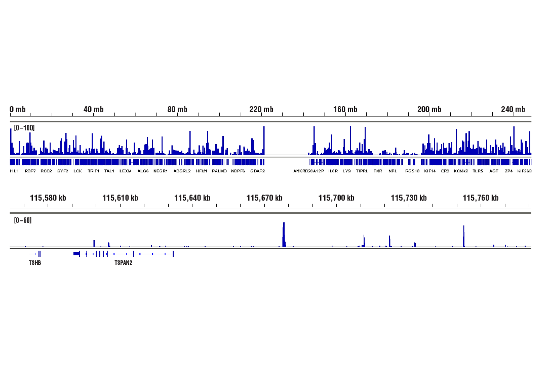
CUT&RUN was performed with NCI-H2052 cells and YAP (D8H1X) XP® Rabbit mAb, using CUT&RUN Assay Kit #86652. DNA libraries were prepared using DNA Library Prep Kit for Illumina® (ChIP-seq, CUT&RUN) #56795. The figures show binding across across chromosome 1 (upper), including TSPAN2 (lower), a known target gene of YAP (see additional figure containing CUT&RUN-qPCR data).