


 下载产品说明书
 用小程序,查商品更便捷


 收藏
收藏
 对比
对比 咨询
咨询
Product Usage Information
For optimal ChIP and ChIP-seq results, use 10 μl of antibody and 10 μg of chromatin (approximately 4 x 10⁶ cells) per IP. This antibody has been validated using SimpleChIP® Enzymatic Chromatin IP Kits.The CUT&RUN dilution was determined using CUT&RUN Assay Kit #86652.
| Application | Dilution |
|---|---|
| Western Blotting | 1:1000 |
| Simple Western™ | 1:10 - 1:50 |
| Immunoprecipitation | 1:50 |
| IHC Leica Bond | 1:100 - 1:400 |
| Immunohistochemistry (Paraffin) | 1:200 - 1:800 |
| Immunofluorescence (Frozen) | 1:50 - 1:200 |
| Immunofluorescence (Immunocytochemistry) | 1:50 - 1:200 |
| Flow Cytometry (Fixed/Permeabilized) | 1:50 - 1:200 |
| Chromatin IP | 1:50 |
| Chromatin IP-seq | 1:50 |
| CUT&RUN | 1:50 |
Specificity/Sensitivity
物种反应性:
人, 小鼠, 大鼠, 仓鼠 , 猴
YAP(Yes 相关蛋白,YAP65)的首次鉴定基于其与 Yes 的 SH3 域结合的能力。它还结合其他包含 SH3 结构域的蛋白,如 Nck、Crk、Src 和 Abl (1)。除了 SH3 结合基序,YAP 还有一个 PDZ 互作基序、一个卷曲螺旋结构域和许多 WW 结构域 (2-4)。YAP 的初步研究都指出,它在锚定和靶向特定亚细胞区室中发挥作用。后续研究表明,YAP 是一个转录共激活蛋白,因为其 WW 结构域与转录因子 PEBP2 和其他转录因子的 PY 基序 (PPxY) 相互作用 (5)。作为一个转录共激活蛋白,YAP 现在被广泛认为是 Hippo 通路的一个中央介导物,从而在组织生长和器官大小的调节中发挥功能性和广泛保守的作用 (6-8)。LATS 激酶磷酸化许多位点(如 Ser109 和 Ser127)会促进 YAP 从细胞核转位到细胞浆,从而通过结合 14-3-3 蛋白被隔离 (7-9)。这些 LATS 诱导的磷酸化活动能让 YAP 进一步被邻近磷酸降解决定子中的 CK1δ/ε 磷酸化,从而诱发 YAP 蛋白酶体降解 (10)。 1.Sudol, M. (1994) Oncogene 9, 2145-52. 2.Mohler, P.J. et al. (1999) J Cell Biol 147, 879-90. 3.Espanel, X. and Sudol, M. (2001) J Biol Chem 276, 14514-23. 4.Sudol, M. et al. (1995) FEBS Lett 369, 67-71. 5.Yagi, R. et al. (1999) EMBO J 18, 2551-62. 6.Dong, J. et al. (2007) Cell 130, 1120-33. 7.Zhao, B. et al. (2010) Genes Dev 24, 862-74. 8.Zhao, B. et al. (2007) Genes Dev 21, 2747-61. 9.Yu, F.X. et al. (2012) Cell 150, 780-91. 10.Zhao, B. et al. (2010) Genes Dev 24, 72-85.
参考图片
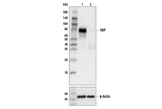
Western blot analysis of extracts from various cell lines using YAP (D8H1X) XP® Rabbit mAb (upper) and β-Actin (D6A8) Rabbit mAb #8457 (lower). As expected, RL-7 cells are negative for YAP protein expression.
Confocal immunofluorescent analysis of low confluence MCF 10A cells (left), high confluence MCF 10A (center), and YAP negative RL-7 cells (right) using YAP (D8H1X) XP® Rabbit mAb (green). Blue pseudocolor in lower images = DRAQ5® #4084 (fluorescent DNA dye). Increased nuclear localization of YAP protein is seen in low confluence (proliferating) cells.
Immunohistochemical analysis of paraffin-embedded PANC-1 (left) and RL-7 (right) cell pellets using YAP (D8H1X) XP® Rabbit mAb.
Immunohistochemical analysis of paraffin-embedded human breast adenocarcinoma using YAP (D8H1X) XP® Rabbit mAb.
Immunoprecipitation of YAP protein from A-204 cell extracts using Rabbit (DA1E) mAb XP® Isotype Control #3900 (lane 2) or YAP (D8H1X) XP® Rabbit mAb (lane 3). Lane 1 is 10% input. Western blot analysis was performed using YAP (D8H1X) XP® Rabbit mAb. Mouse Anti-rabbit IgG (Conformation Specific) (L27A9) mAb #3678 was used as a secondary antibody.
Flow cytometric analysis of RL-7 cells (blue) and A-204 cells (green) using YAP (D8H1X) XP® Rabbit mAb. Anti-rabbit IgG (H+L), F(ab')2 Fragment (Alexa Fluor® 488 Conjugate) #4412 was used as a secondary antibody.
Immunohistochemical analysis of paraffin-embedded human breast adenocarcinoma using YAP (D8H1X) XP® Rabbit mAb.
Immunohistochemical analysis of paraffin-embedded human ovarian serous carcinoma using YAP (D8H1X) XP® Rabbit mAb.
Immunohistochemical analysis of paraffin-embedded human benign prostatic hyperplasia using YAP (D8H1X) XP® Rabbit mAb.
Chromatin immunoprecipitations were performed with cross-linked chromatin from 4 x 10⁶ NCI-2052 cells and either 10 µl of YAP (D8H1X) XP® Rabbit mAb, or 2 µl of Normal Rabbit IgG #2729, using SimpleChIP® Enzymatic Chromatin IP Kit (Magnetic Beads) #9003. The enriched DNA was quantified by real-time PCR using SimpleChIP® Human CTGF Promoter Primers #14927, human SMYD3 intron 2 primers, and SimpleChIP® Human CTGF Upstream Primers #14928. The amount of immunoprecipitated DNA in each sample is represented as signal relative to the total amount of input chromatin, which is equivalent to one.

 危险品化学品经营许可证(不带存储) 许可证编号:沪(杨)应急管危经许[2022]202944(QY)
  营业执照(三证合一)